Table of Contents
Overview – Urinary System Anatomy
The urinary system anatomy underpins many key physiological functions in the human body, including waste excretion, fluid balance, electrolyte regulation, and blood pressure homeostasis. Understanding the structural organisation of the urinary system is essential for clinical reasoning in renal and systemic pathology. This article will explore both the macroscopic and microscopic anatomy of the kidneys, ureters, bladder, and urethra, highlighting their functional roles and clinical relevance.
Definition
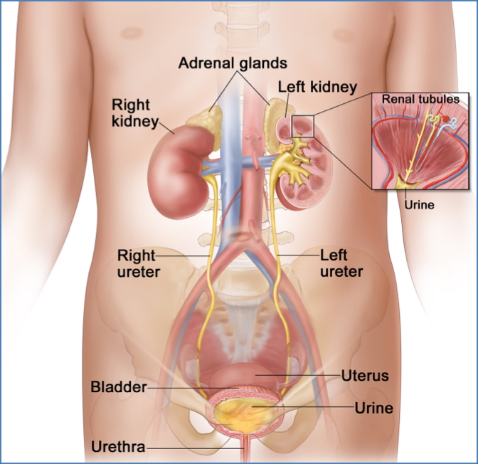
The urinary system comprises organs that filter the blood, produce urine, and eliminate waste products. It includes the kidneys, ureters, bladder, and urethra. These structures work together to maintain internal fluid and electrolyte balance and to excrete metabolic waste products via urine.
Functions of the Urinary System
- Ultrafiltration of blood (removal of particles, bacteria, viruses)
- Excretion of metabolic waste and drugs
- Regulation of:
- Water and electrolyte balance
- Body fluid osmolality
- Blood volume and arterial pressure
- Acid-base balance (with respiratory system)
- Storage and elimination of urine
- Endocrine secretion (e.g. renin, erythropoietin)
- Gluconeogenesis (e.g. from amino acids)
- Male reproductive function (urethral integration)
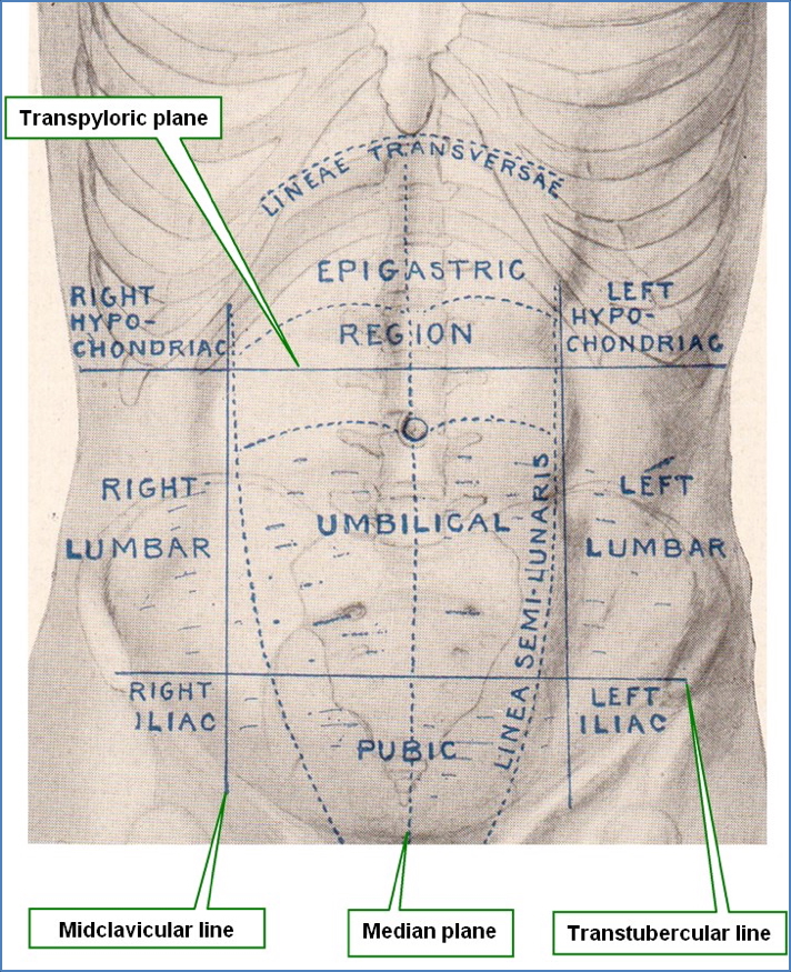
Surface Anatomy of the Kidneys
- Level: Transpyloric plane (T12) crosses the hilum of the left kidney and superior pole of the right kidney
- Midline relations: Hila ~5 cm from midline, diverging slightly at inferior poles
- Height & position:
- Posterior to ribs 11–12
- Move ~2–3 cm with respiration
- Right kidney lies slightly lower than left (~2.5 cm) due to liver
- Dimensions:
- 12 cm long, 5–6 cm wide, 3–4 cm thick
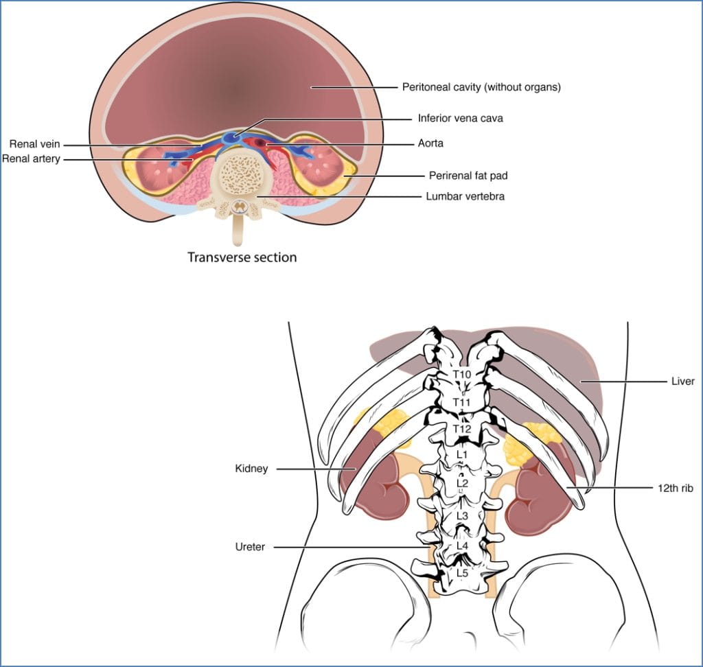
Anatomical Relationships
- Retroperitoneal organs
- Surrounding structures:
- Left kidney: spleen (lateral), pancreas (anterior), descending colon
- Right kidney: liver (superior), duodenum & ascending colon (anterior)
- Adrenal glands: superior poles of both kidneys
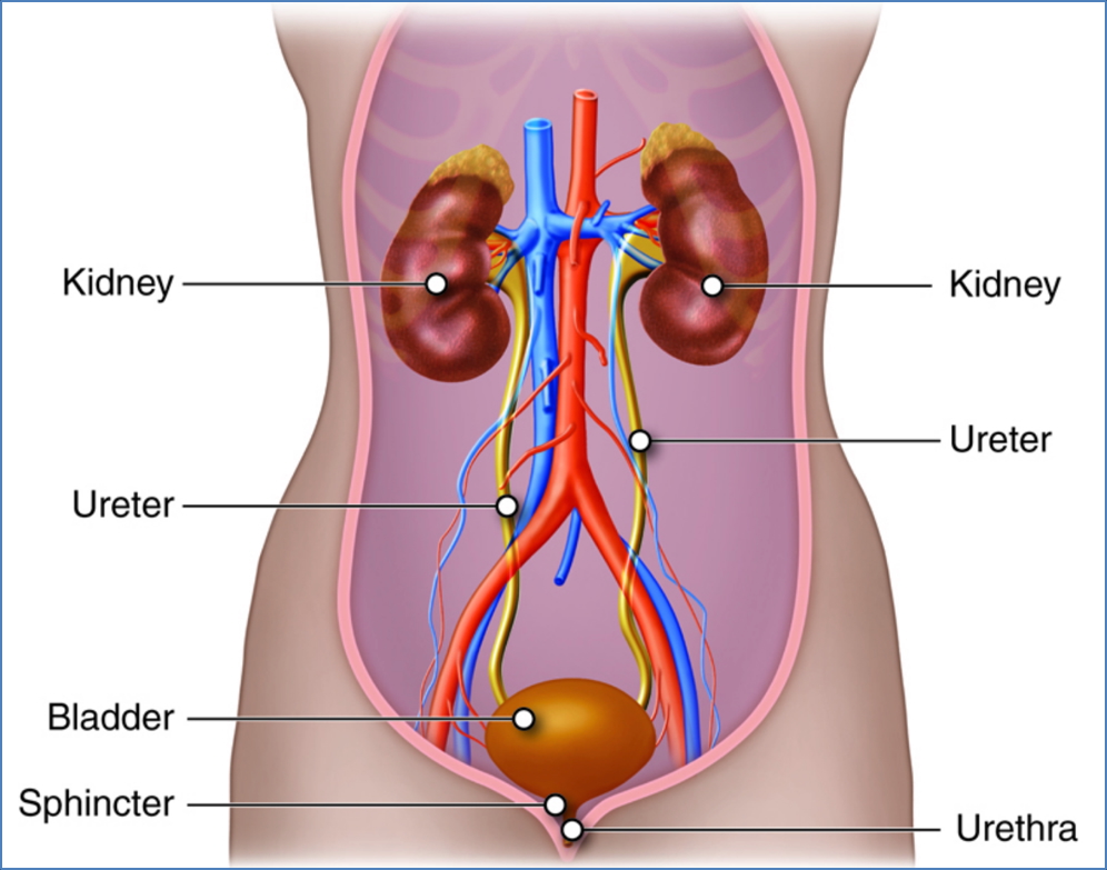
Functional Components of the Urinary System
- Kidneys: Filter blood, produce urine, maintain homeostasis
- Renal arteries/veins: Enter/exit at hilum, supply and drain kidneys
- Ureters: Transport urine to bladder
- Bladder: Stores urine until micturition
- Urethra: Conducts urine outside the body

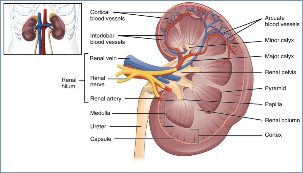
Macroscopic Kidney Anatomy
- Protective structures: Renal capsule, perinephric fat, renal fascia
- Cortex: Contains renal corpuscles, blood vessels, proximal/distal tubules
- Medulla: Renal pyramids and columns, collecting ducts
- Collecting system:
- Renal papilla → minor calyces → major calyces → renal pelvis → ureter
- Lobes: Pyramid + overlying cortex
- Hilum: Entry/exit point for vessels and ureter
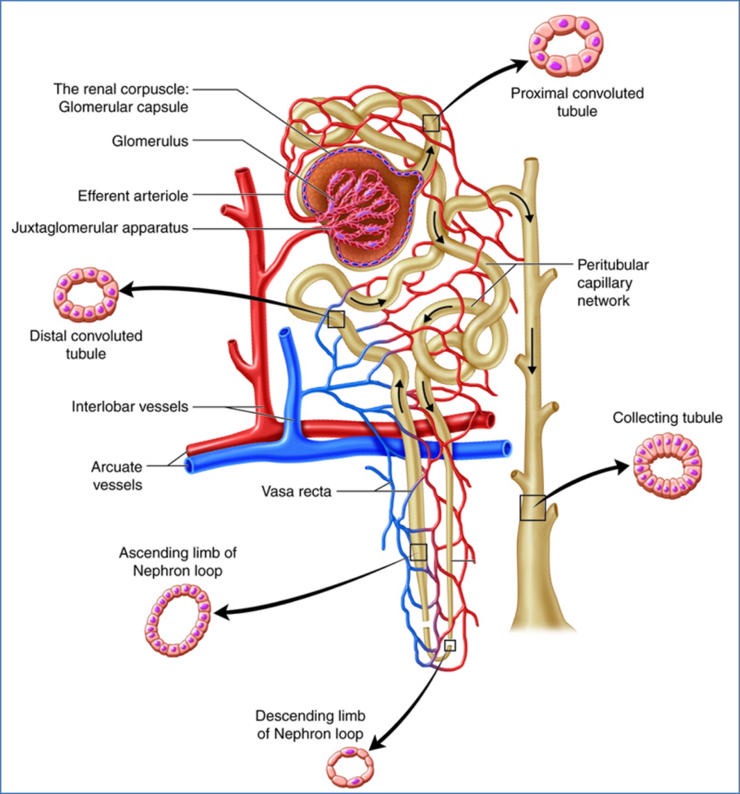
Microscopic Kidney Anatomy
- Microvasculature:
- Interlobar arteries → arcuate arteries → interlobular arteries → afferent arterioles
- Efferent arterioles → peritubular capillaries → venules → renal vein → IVC
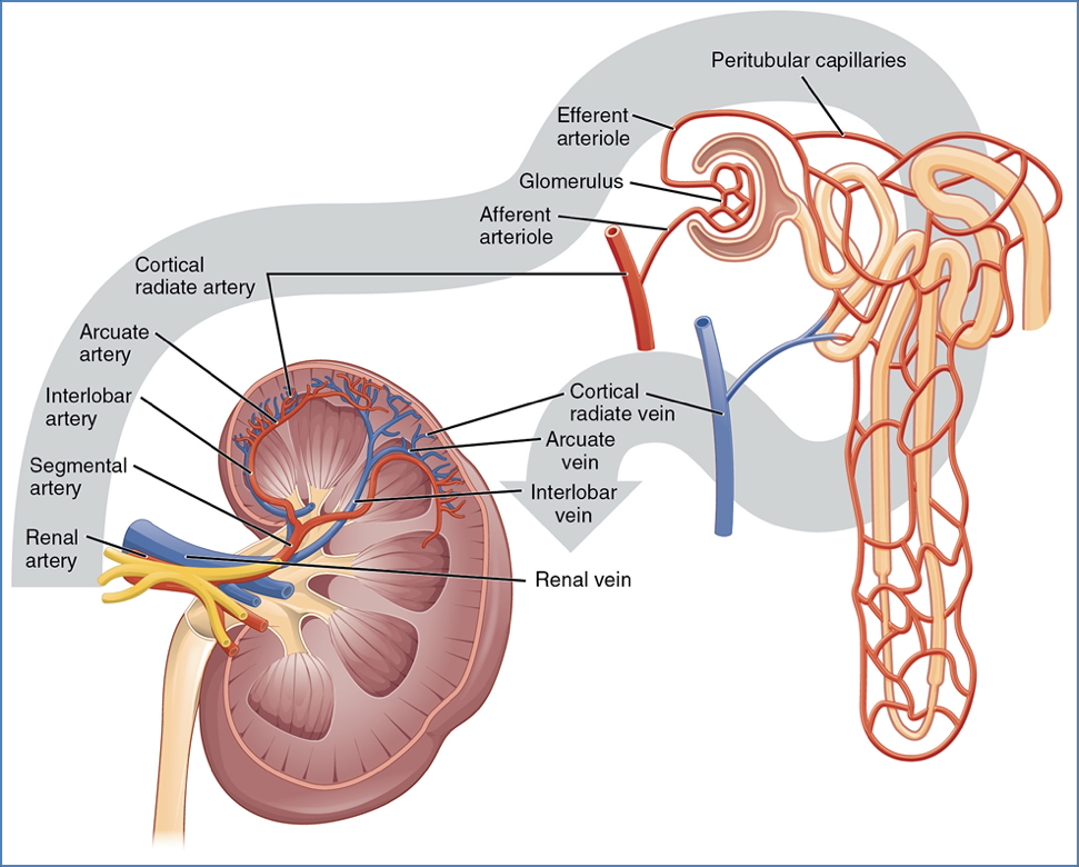
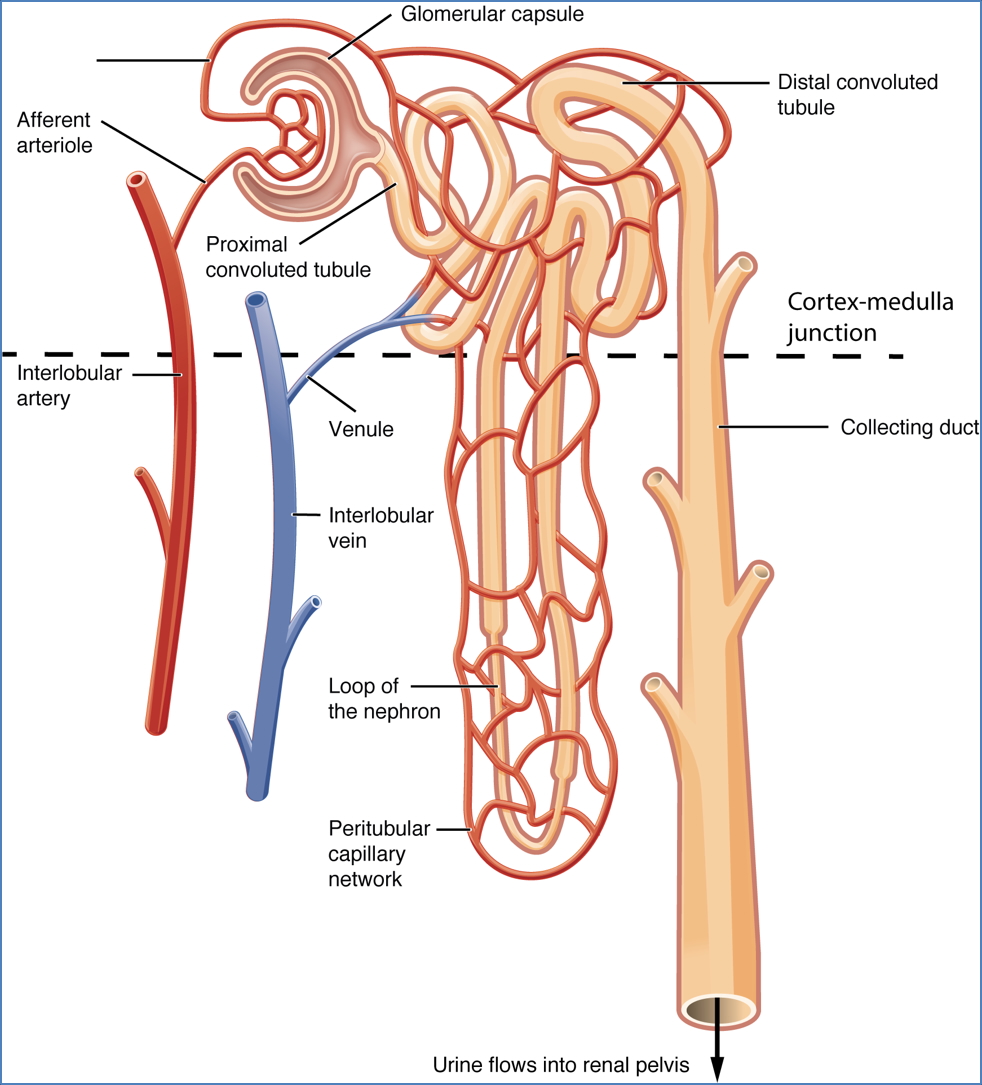
- Renal Corpuscle:
- Glomerulus + Bowman’s capsule
- Site of blood filtration
- Renal Tubule Components:
- Proximal Convoluted Tubule: Reabsorbs water, ions, nutrients
- Loop of Henle:
- Descending limb: water reabsorption (simple squamous epithelium)
- Ascending limb: ion reabsorption (cuboidal epithelium)
- Distal Convoluted Tubule: Variable Na⁺, Ca²⁺, water reabsorption; secretion of wastes
- Collecting duct system: Final urine modification before excretion

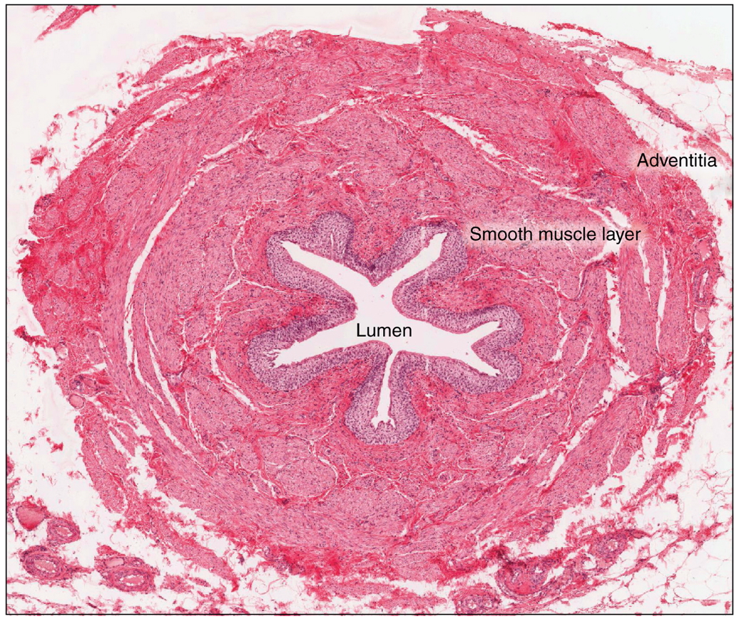
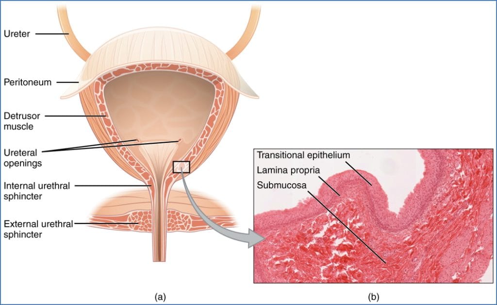
Ureters
- Length: ~30–35 cm
- Structure: Muscular tubes with transitional epithelium lining
- Course:
- Abdominal segment anterior to psoas major
- Pelvic segment below bifurcation of common iliac artery
- Constriction points:
- Ureteropelvic junction
- Pelvic brim
- Vesicoureteric junction
- Blood supply:
- Upper: renal artery branches
- Middle: gonadal, aortic, common iliac branches
- Lower: internal iliac artery branches
2. https://www.ncbi.nlm.nih.gov/books/NBK65953/
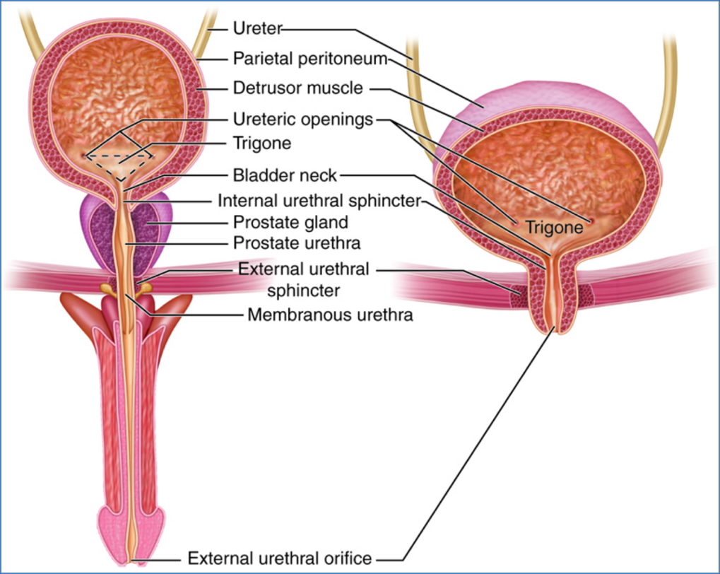
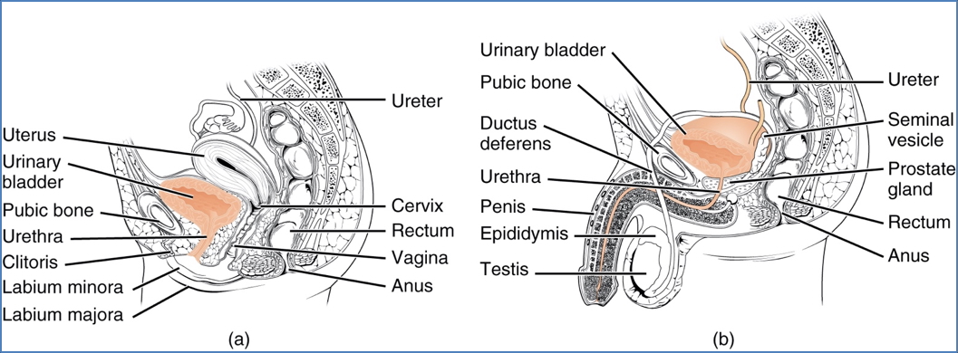
Bladder
- Location: Inferior to peritoneum; posterior to pubic symphysis
- Regions: Apex, neck, body, fundus, trigone
- Trigone: Smooth triangular area defined by ureteric and urethral orifices
- Histology:
- Transitional epithelium
- Detrusor muscle
- Gender-specific anatomy:
- Male: Rectovesical pouch behind bladder
- Female: Vesicouterine pouch between bladder and uterus
- Blood supply: Internal iliac artery (± vaginal artery in females)
Urethra
- Function: Excretes urine from bladder
- Sphincters:
- Internal (involuntary): at bladder neck
- External (voluntary): within pelvic diaphragm
- Length:
- Male: ~20 cm (prostatic, membranous, spongy sections)
- Female: ~2–3 cm
- Histology:
- Male: transitional → pseudostratified columnar
- Female: pseudostratified columnar → stratified squamous (external)
2. OpenStax College, CC BY 3.0 <https://creativecommons.org/licenses/by/3.0>, via Wikimedia Commons
Summary – Urinary System Anatomy
The urinary system anatomy encompasses multiple structures involved in blood filtration, urine production, and excretion. From the kidneys’ intricate microvasculature and nephron architecture to the functional dynamics of ureters, bladder, and urethra, each component plays a crucial role in maintaining homeostasis. A clear grasp of urinary system anatomy forms the foundation for understanding renal physiology and pathology. For a broader context, see our Renal Overview page.