Table of Contents
Overview – Dermatology Basics
A strong understanding of dermatology basics—including lesion terminology, morphology, distribution, and classification—is essential for accurate clinical diagnosis. Whether it’s differentiating a macule from a papule or recognising the patterns of erythema versus ulceration, final-year medical students must be confident with dermatological language. This guide to dermatology basics outlines the classification of primary and secondary skin lesions, colour descriptors, and other key clinical terms used in dermatological assessment.
Primary vs Secondary Lesions
Primary Lesions
- Originate directly from the disease process
- Serve as the initial identifying feature of a skin condition
Secondary Lesions
- Develop from progression, modification, or trauma to a primary lesion
- May involve healing, infection, or scratching
Primary Lesion Terminology
| Lesion Name | Description | Example Image |
|---|---|---|
| Erythema | Redness due to vascular dilation (e.g. cellulitis) | 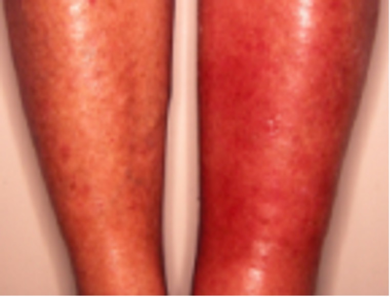 |
| Erythroderma | Generalised erythema covering >90% of the body |  |
| Telangiectasia | Visibly dilated dermal blood vessels; blanchable | 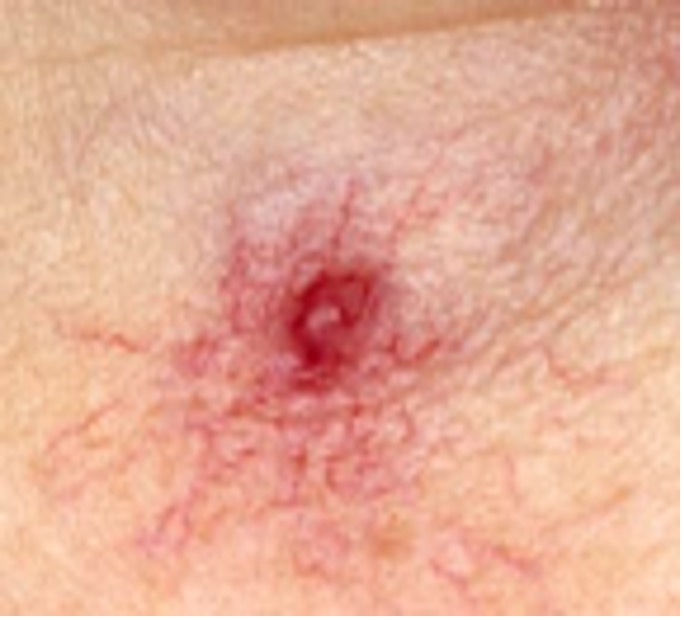 |
| Macule | Flat, non-palpable area of altered colour <1 cm (e.g. freckles) | 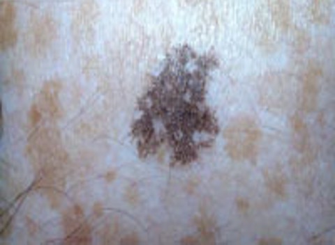 |
| Patch | Larger version of a macule |  |
| Petechiae | Tiny, non-blanching macules due to RBC extravasation |  |
| Purpura | Larger, non-blanching macule/patch of blood in the skin | 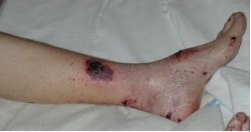 |
| Ecchymosis | Large subcutaneous bleed; may form a haematoma | 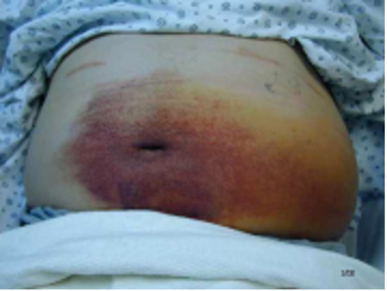 |
| Micropapule | Tiny raised lesion (1–2 mm) |  |
| Papule | Small, raised lesion <0.5 cm |  |
| Plaque | Flat-topped raised lesion >2 cm width, no depth (e.g. psoriasis) |  |
| Papilloma | Nipple-like skin projection | 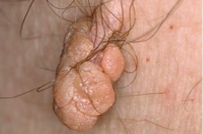 |
| Burrow | Linear papule due to burrowing organism (e.g. scabies) |  |
| Nodule | Raised, solid lesion >0.5 cm width and depth |  |
| Tumour | Solid mass >1 cm |  |
| Vesicle | Clear, fluid-filled blister <5 mm |  |
| Bulla | Larger fluid-filled lesion >0.5 cm | 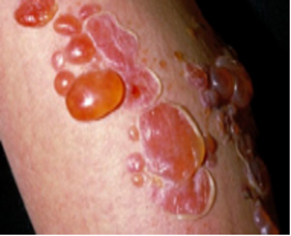 |
| Pustule | Pus-filled vesicle <5 mm | 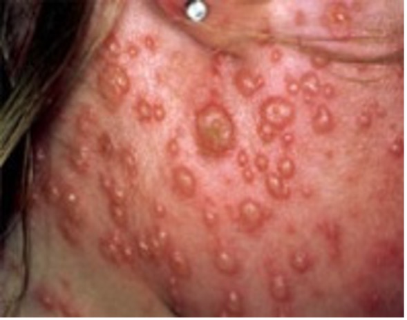 |
| Impetiginised | Crusted, pustular, weeping lesion typical of impetigo | 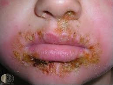 |
| Cyst | Cavity containing fluid, semisolid or solid material |  |
| Abscess | Pus-filled cavity >1 cm; may result from infected cyst |  |
| Wheal | Transient, white, compressible swelling from dermal oedema |  |
| Angioedema | Oedema extending into the subcutaneous tissue |  |
| Comedone | Keratin/sebum plug (e.g. blackhead) |  |
| Alopecia | Hair loss: can be scarring or non-scarring |  |
| Eschar | Black necrotic tissue over ulcer or erosion; indicates severe vascular compromise | 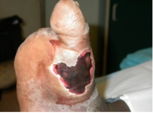 |
Secondary Lesion Terminology
| Lesion Name | Description | Example Image |
|---|---|---|
| Scale | Dry masses of keratin (e.g. psoriasis) |  |
| Keratosis | Skin thickening (e.g. actinic keratosis) |  |
| Hyperkeratosis | Generalised thickening of the stratum corneum | 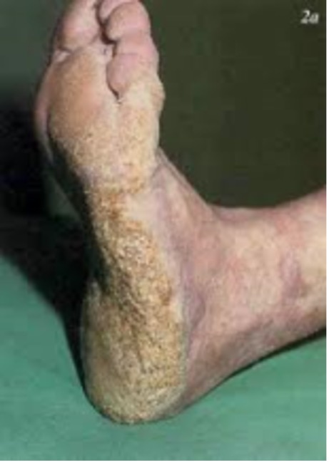 |
| Verrucous | Warty, hyperkeratotic lesion |  |
| Lichenification | Palpable thickening from repeated friction (e.g. lichen simplex) |  |
| Crust | Dried serum, pus, or blood over damaged epithelium | 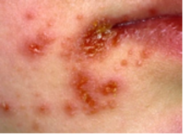 |
| Atrophy | Thinning of the epidermis, dermis, or subcutaneous fat | 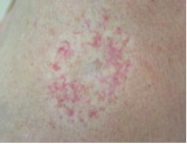 |
| Erosion | Superficial partial epidermal loss (e.g. superficial burn) | 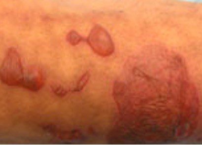 |
| Ulcer | Full-thickness skin loss extending to dermis or fat |  |
| Excoriation | Linear epidermal loss due to scratching |  |
| Scar | Raised, firm or depressed lesion post-injury (e.g. keloid) |  |
| Striae | Stretch marks; linear bands of altered connective tissue |  |
| Pigmentation | Hyper- or hypopigmented changes (e.g. birthmarks) |  |
Descriptive Terms in Dermatology
Colour
- Erythema – Red
- Violaceous – Purple
- Slate – Grey
- Hyperpigmented – Darker than surrounding skin
- Hypopigmented – Lighter than surrounding skin
- Bronze – Reddish-brown
- Dusky – Purple-grey
- Variegated – Multi-coloured
Edge Definition & Shape
- Defined – Clear edge
- Ill-defined – Blurred margin
- Nummular – Round
- Annular – Ring-like
- Circinate – Circular
- Arcuate – Curved
- Discoid – Disc-shaped
- Serpiginous/Gyrate – Wavy or meandering
- Polycyclic – Overlapping circular shapes
Surface Countours

Surface Features & Sensory Descriptions
- Weeping – Oozing clear fluid
- Crusted – Covered with dried exudate
- Pruritic – Itchy
- Dysaesthesia – Burning, tingling, or numbness
Distribution & Symmetry
- Symmetrical Lesions:
- Suggest immune/allergic causes (e.g. eczema, psoriasis)
- Unilateral or Localised Lesions:
- Suggest infection or local trauma (e.g. abscess, shingles)
Typical Distributions
- Seborrhoeic Dermatitis – Scalp, forehead, eyebrows, chest
- Atopic Dermatitis – Cubital/popliteal fossae
Extent
- Localised – Confined area (e.g. shingles)
- Universal – Generalised over body (e.g. chickenpox)
Summary – Dermatology Basics
Mastering dermatology basics allows for accurate recognition and communication of skin findings, from primary lesions like macules and papules to secondary changes such as scaling, crusting, or ulceration. Colour, symmetry, and distribution also provide critical diagnostic clues. This comprehensive reference on dermatology basics supports clinical reasoning and documentation in dermatology. For a broader context, see our Skin & Dermatology Overview page.