Table of Contents
Overview – Gastrointestinal System
The gastrointestinal (GI) system is central to digestion, nutrient absorption, and waste elimination. It encompasses a complex network of organs and accessory structures that work in harmony to mechanically and chemically break down food, absorb nutrients, and excrete waste. Understanding the gastrointestinal system is essential for final-year medical students as it underpins the pathophysiology of many acute and chronic conditions encountered in clinical settings.
Definition
The gastrointestinal system, also known as the digestive tract, is a continuous hollow tube extending from the mouth to the anus, including both the alimentary canal and accessory organs. It supports ingestion, digestion, absorption, and excretion.
General Functions of the GI Tract
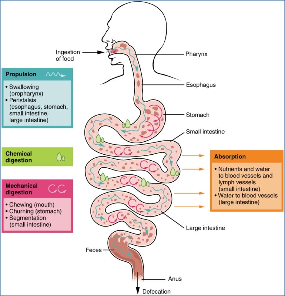
- Ingestion: Entry of food via the mouth.
- Mechanical digestion: Chewing, churning, segmentation, and peristalsis.
- Propulsion: Swallowing and movement through the intestines.
- Chemical digestion: Enzymatic and acidic breakdown of food into absorbable molecules.
- Secretion: Release of enzymes, acids, mucus, and bile.
- Absorption: Uptake of nutrients and fluids into blood and lymph.
- Excretion/Defecation: Elimination of indigestible substances and waste.

Structural Overview
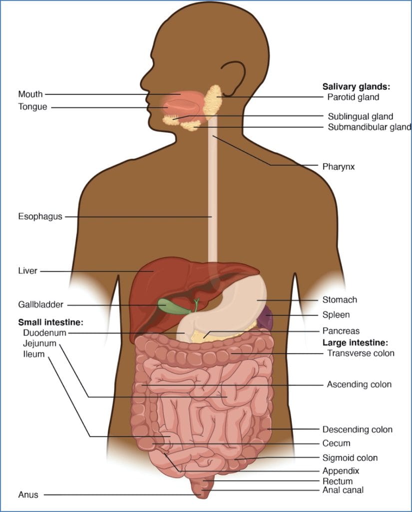
Alimentary Canal (~9 metres)
- Mouth → Mechanical digestion and saliva secretion
- Pharynx → Pathway for food from mouth to oesophagus
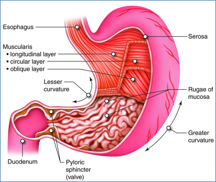
- Oesophagus
- ~25 cm long
- Stratified squamous epithelium
- Upper 1/3: Striated muscle; Lower 2/3: Smooth muscle
- Stomach → Acid and enzyme breakdown; limited absorption
- Small Intestine
- Duodenum, Jejunum, Ileum
- Major site for digestion and absorption
- Large Intestine
- Cecum, Colon, Rectum
- Absorbs water, ions, vitamins; compacts waste
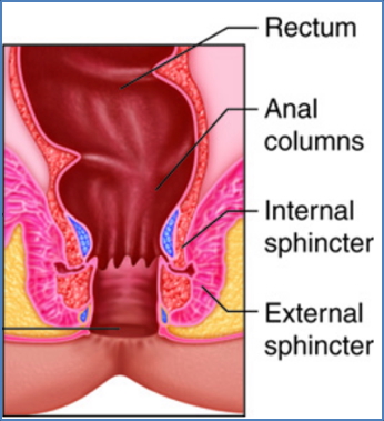
- Anus → Defecation
Accessory Digestive Organs
- Teeth & Tongue → Mastication and food manipulation
- Salivary Glands: Parotid, Sublingual, Submandibular
- Liver → Bile production
- Gallbladder → Bile storage and concentration
- Pancreas → Secretes digestive enzymes and bicarbonate
Specialised Structures
Sphincters
- Upper and Lower Oesophageal
- Pyloric
- Ileocecal
- Internal and External Anal

Pacemaker Zones
- Control peristalsis rhythm:
- Stomach: ~3 contractions/min
- Duodenum: 9–12/min
- Colon: ~2/hour
Temporary Storage Sites
- Mouth, Stomach, Colon, Rectum
Absorptive Enhancers
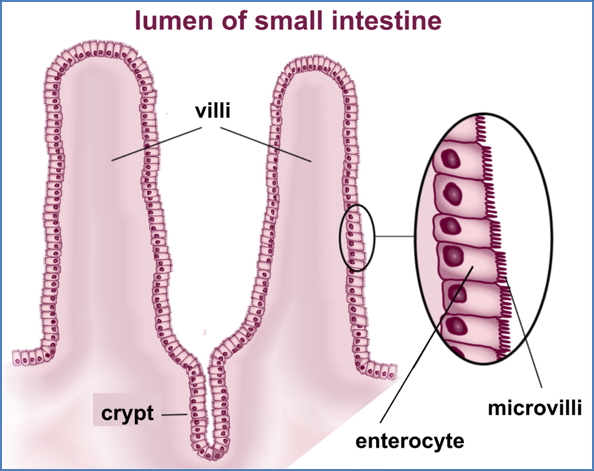
- Plicae circulares, Villi, Microvilli: Increase surface area for absorption
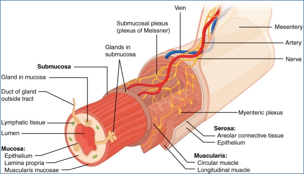
Histological Layers of the Gastrointestinal Tract
1. Mucosa
- Histology:
- Epithelium (simple columnar with goblet cells)
- Lamina propria (areolar tissue)
- Muscularis mucosae (smooth muscle)
- Functions:
- Secretes enzymes, mucus, hormones
- Absorbs nutrients
- Protects from acid, bacteria, mechanical damage
2. Submucosa
- Dense connective tissue with vessels, glands, nerves
- Supports mucosa and allows elasticity
3. Muscularis
- Inner circular and outer longitudinal smooth muscle layers
- Drives peristalsis and forms sphincters
4. Serosa (Visceral Peritoneum)
- Areolar connective tissue + mesothelium
- Provides lubrication and structural support
Digestion Phases and Enzymes
Mechanical Digestion
- Chewing (oral cavity)
- Churning (stomach)
- Segmental mixing (intestines)
Chemical Digestion
- Salivary Amylase → Carbohydrate digestion (mouth)
- Pepsin + HCl → Protein digestion (stomach)
- Bile (liver) → Emulsification of fats
- Pancreatic enzymes:
- Amylase (carbs)
- Lipase (fats)
- Proteases (proteins)
- Nucleases (DNA/RNA)
Intestinal Absorption
- Blood vessels: Absorb water, carbs, amino acids, vitamins
- Lymphatics: Absorb fats and fat-soluble vitamins
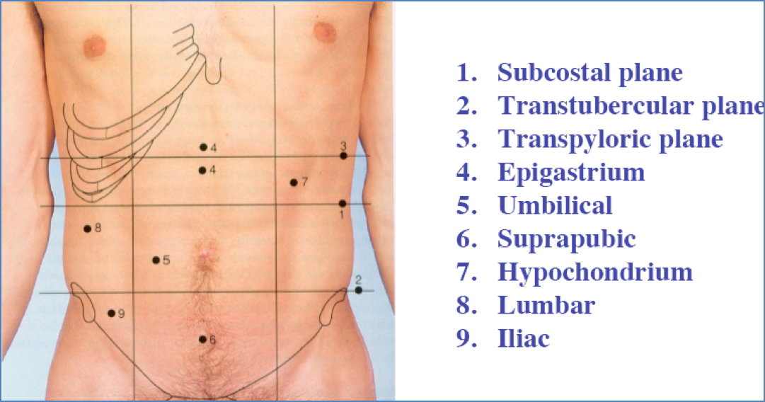
Abdominal Cavity
Boundaries
- Superior: Thoracic diaphragm
- Inferior: Broad ligament of pelvis


Wall Layers (External to Internal)
- Skin → Superficial fascia (fatty & membranous)
- External oblique
- Internal oblique
- Transverse abdominal muscle
- Transversalis fascia
- Parietal peritoneum

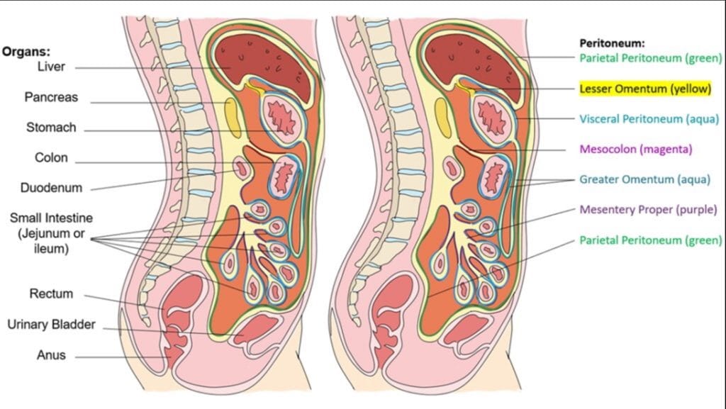
The Peritoneum
Layers
- Visceral peritoneum: Covers digestive organs
- Parietal peritoneum: Lines abdominal wall
- Peritoneal cavity: Contains lubricating serous fluid
Mesenteries
- Double layers of peritoneum anchoring organs
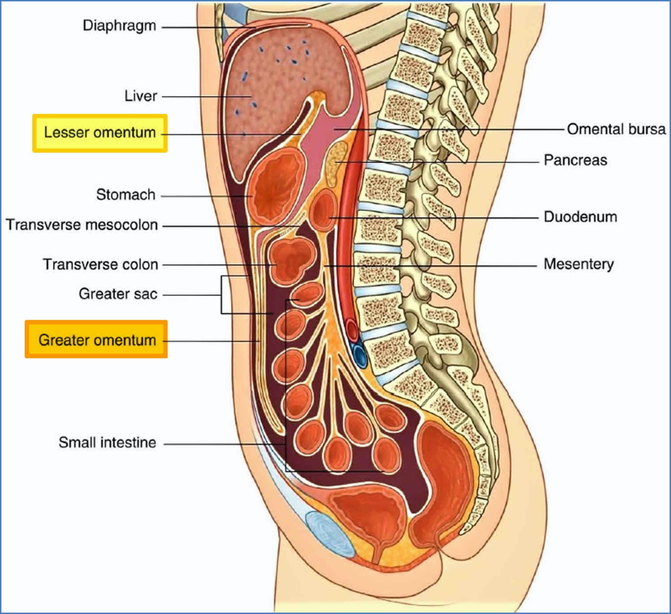
- Lesser omentum: Lesser curvature of stomach
- Greater omentum: Greater curvature
- Mesocolon: Colon
- Mesentery proper: Jejunum, ileum
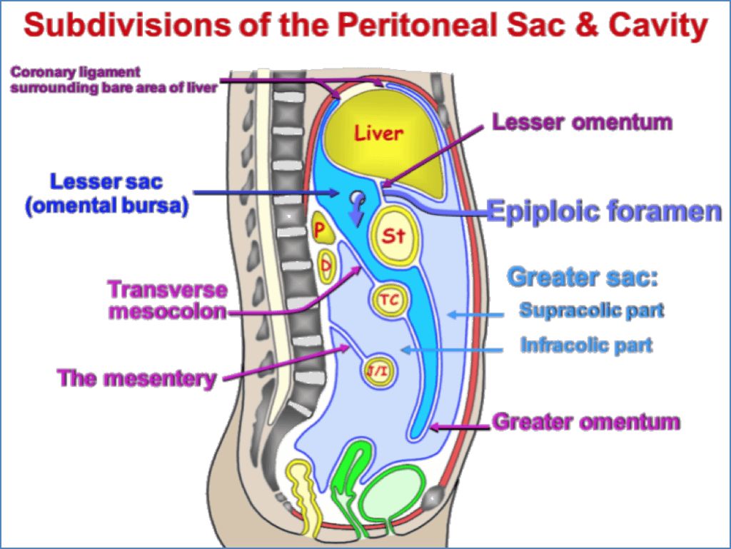
Subdivisions
- Greater sac: Supracolic & infracolic compartments
- Lesser sac (omental bursa): Posterior to stomach
Intra- vs Retroperitoneal Organs
- Intra-peritoneal:
- Stomach, Gallbladder, Jejunum, Ileum, Cecum, Transverse & Sigmoid colon
- Retroperitoneal:
- Duodenum (parts), Pancreas (majority), Ascending/Descending colon, Rectum
Summary – Gastrointestinal System
The gastrointestinal system is essential for processing food into nutrients, eliminating waste, and maintaining internal homeostasis. It comprises the alimentary canal and accessory organs, with specialised histological layers, digestive enzymes, and peritoneal structures contributing to its function. Understanding the gastrointestinal system is foundational for clinical reasoning in medicine. For a broader context, see our Gastrointestinal Overview page.