Table of Contents
Overview – The Vascular System
The vascular system comprises arteries, capillaries, and veins that work together to circulate blood throughout the body. Each component of the system has a distinct structure and function, from the high-pressure conduits of elastic arteries to the exchange-focused capillaries and the capacitance-based return pathway of veins. Understanding vascular anatomy and physiology is critical for diagnosing and managing cardiovascular conditions in clinical practice.
The Arterial System
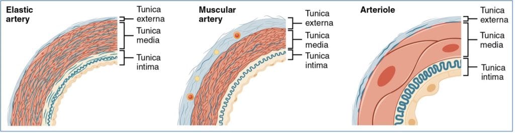
Elastic (Conducting) Arteries
- Includes the aorta and its major branches
- Thick-walled with large lumens → low resistance to flow
- High elastin content allows:
- Dampening of pressure fluctuations
- Energy storage during systole → blood propulsion during diastole
Muscular (Distributing) Arteries
- Distal to elastic arteries; supply specific organs
- Diameter: 0.3 mm to 1 cm
- Thickest tunica media with high smooth muscle content
- Highly active in vasoconstriction → less elastic
- Key regulators of regional blood distribution
Arterioles
- Smallest arteries
- Larger arterioles have all 3 tunics (intima, media, externa)
- Smaller arterioles = 1–2 layers of smooth muscle surrounding endothelium
- Critical for local blood flow control
- Respond to:
- Neural signals (e.g. sympathetic tone)
- Hormones (e.g. norepinephrine, vasopressin)
- Local chemicals (e.g. NO, endothelin)
- Vasoconstriction → bypasses capillary beds
- Vasodilation → allows tissue perfusion
- Respond to:
- Major determinant of systemic blood pressure
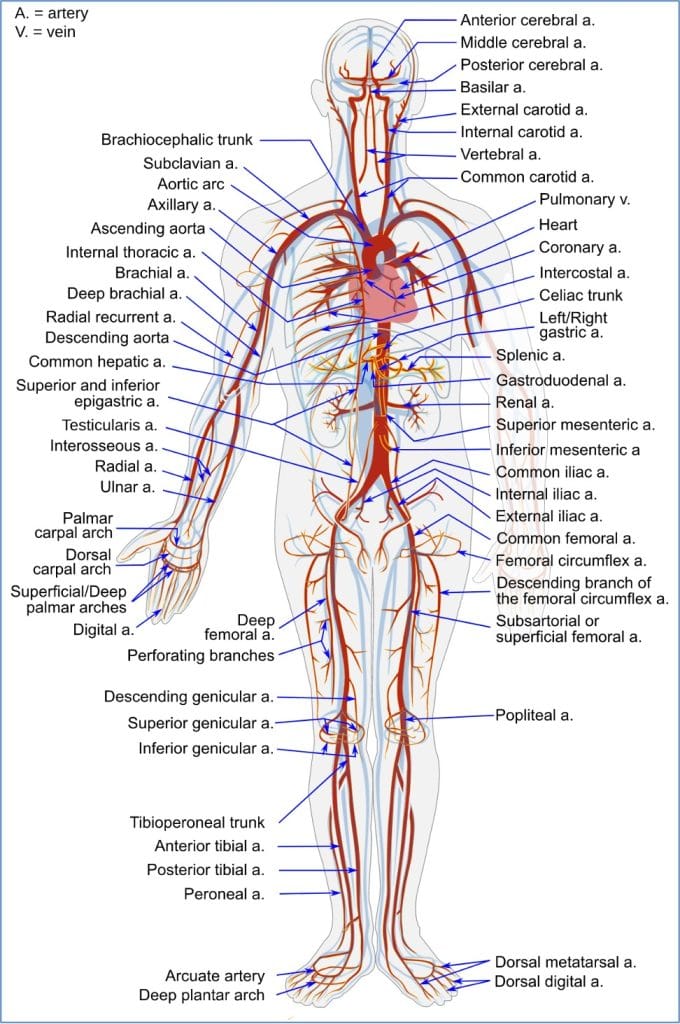
Arterial System Anatomy
The Capillary System
Structure and Function
- Microscopic vessels with thin walls (only tunica intima)
- Length ≈ 1 mm; diameter just enough to allow RBCs in single file
- Site of exchange between blood and interstitial fluid
- Penetrate most tissues except cartilage, tendons, ligaments, epithelia
Capillary Beds
- Network of capillaries = site of microcirculation
- Blood flow from arteriole → venule
- Components:
- Vascular Shunt:
- Metarteriole to thoroughfare channel
- Direct arteriole-to-venule connection
- True Capillaries:
- Branch from metarteriole
- Perform exchange functions
- Vascular Shunt:
- Precapillary Sphincters:
- Smooth muscle cuffs at capillary roots
- Regulate entry of blood into true capillaries
- Allow blood to bypass or enter capillary beds based on tissue demand

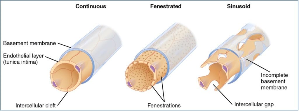
Types of Capillaries
- Continuous Capillaries:
- Uninterrupted endothelium with tight junctions
- Intercellular clefts allow limited solute passage
- Form blood-brain barrier in CNS
- Fenestrated Capillaries:
- Contain fenestrations (pores) → ↑ permeability
- Found in kidneys, intestines, endocrine organs
- Sinusoids (Sinusoidal Capillaries):
- Large, irregular lumens
- Incomplete basement membranes
- Found in liver, bone marrow, lymphoid tissue
- Allow passage of large molecules and WBCs
- Lined with phagocytic Kupffer cells
The Venous System
Venules
- Formed from post-capillary capillary union
- Composed of endothelium (high permeability)
- Permit WBC migration into inflamed tissues
- Larger venules:
- Contain smooth muscle (tunica media)
- Thin tunica externa
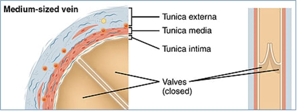
Veins
- Formed from the convergence of venules
- Thinner walls and larger lumens than arteries
- All 3 tunics present, but tunica media poorly developed
- Tunica externa is the thickest layer → collagen & elastic fibers
- Serve as capacitance vessels → hold ~65% of total blood volume
- Low-pressure system requiring adaptations:
- Valves (from tunica intima):
- Prevent backflow
- Function similar to semilunar valves
- Most important in limbs (against gravity)
- Failure can lead to varicosities and thrombosis
- Valves (from tunica intima):
Venous System Anatomy

Foetal Circulation
Shunts Bypassing Certain Organs
- Ductus Venosus:
- Bypasses liver sinusoids
- Directs blood from placenta → inferior vena cava
- Foramen Ovale:
- Interatrial septal opening
- Allows blood to bypass non-functional fetal lungs
- Ductus Arteriosus:
- Connects pulmonary trunk to aorta
- Also bypasses lungs
All fetal shunts normally close at birth due to circulatory pressure changes
- Foramen ovale may take up to 6 months to fully seal

Summary – The Vascular System
The vascular system comprises a complex network of arteries, capillaries, and veins, each uniquely structured to fulfill transport and exchange roles. Understanding these vascular segments, from high-pressure elastic arteries to low-pressure veins, is critical for mastering cardiovascular physiology. For a broader context, see our Cardiovascular Overview page.