Table of Contents
Overview – Blood Vessel Physiology
Blood vessel physiology is central to understanding how blood is transported, exchanged, and regulated throughout the cardiovascular system. Blood vessels function not only as passive conduits but also as dynamic regulators of pressure, flow, and fluid exchange, adjusting to metabolic needs, injury, and systemic changes.
Classification of Blood Vessels
Arteries – Carry blood away from the heart
- Elastic Arteries (Conducting vessels)
- E.g. Aorta and major branches
- Muscular Arteries (Distributing vessels)
- E.g. Coeliac trunk, renal arteries
- Arterioles (Resistance vessels)
- E.g. Intra-organ arterioles
- Terminal Arterioles
- E.g. Afferent arteriole in the kidney
Capillaries – Exchange vessels
- Enable direct contact with tissue for nutrient and waste exchange
- Types:
- Vascular Shunts
- True Capillaries
Veins – Return blood to the heart
- Post-Capillary Venules
- Formed by the union of capillaries
- Small and Large Veins
- Capacitance vessels (contain ~65% of total blood volume)

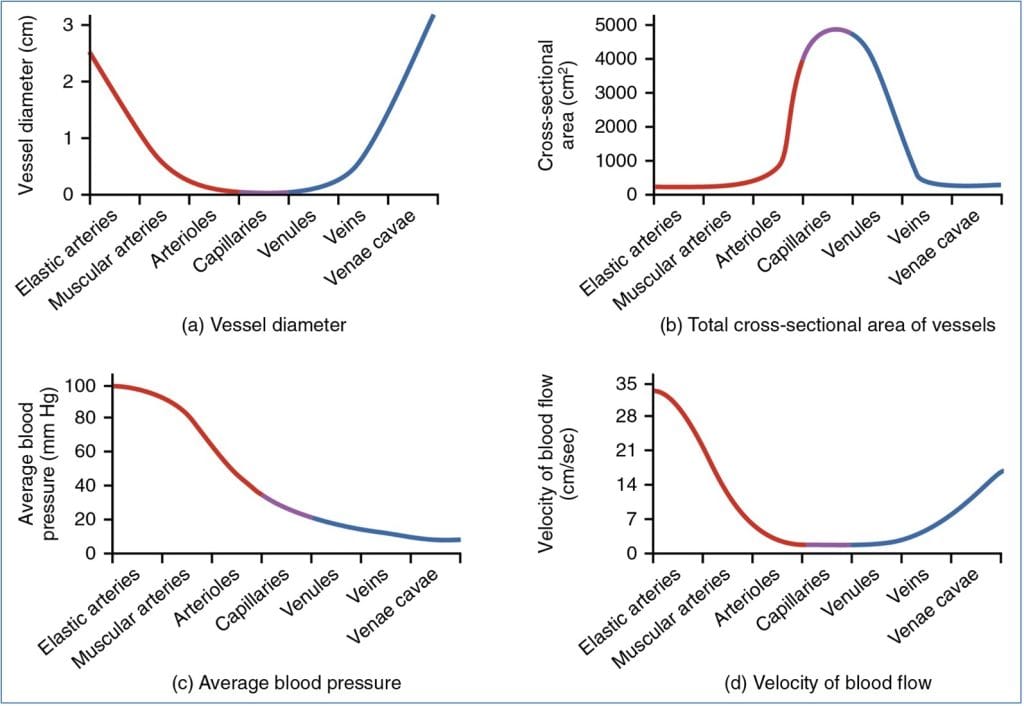
Relationships Between Vessel Diameter, Cross Sectional Area, Local Blood Pressure & Velocity of Flow:
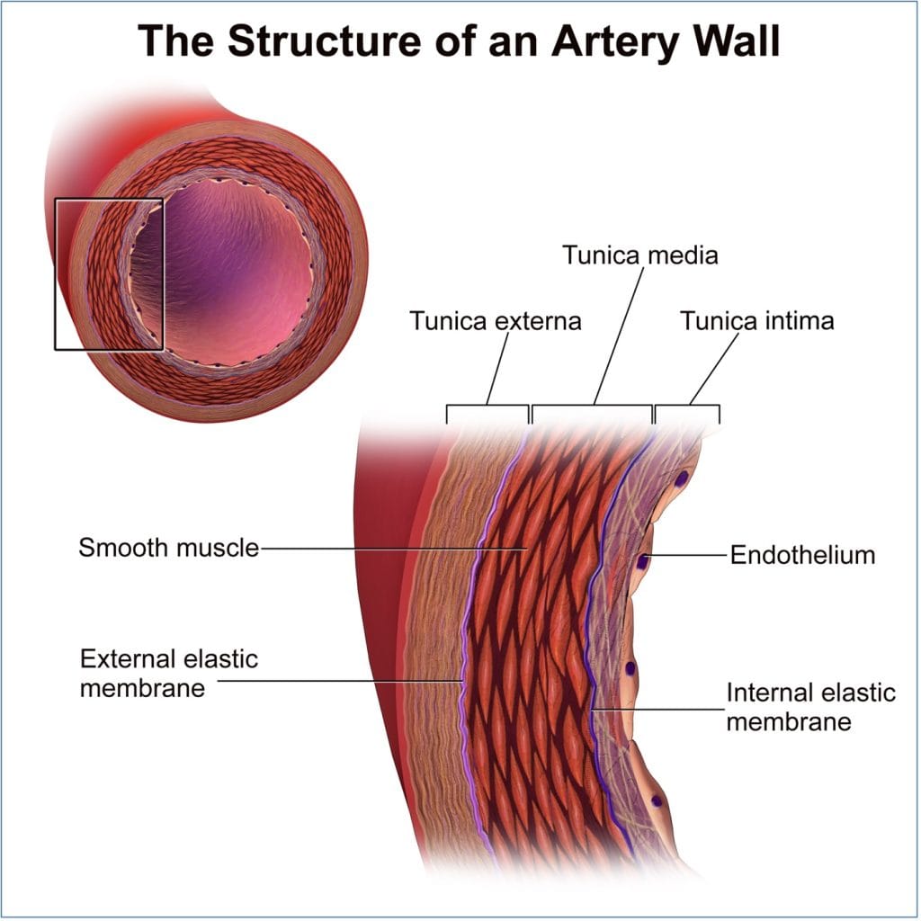
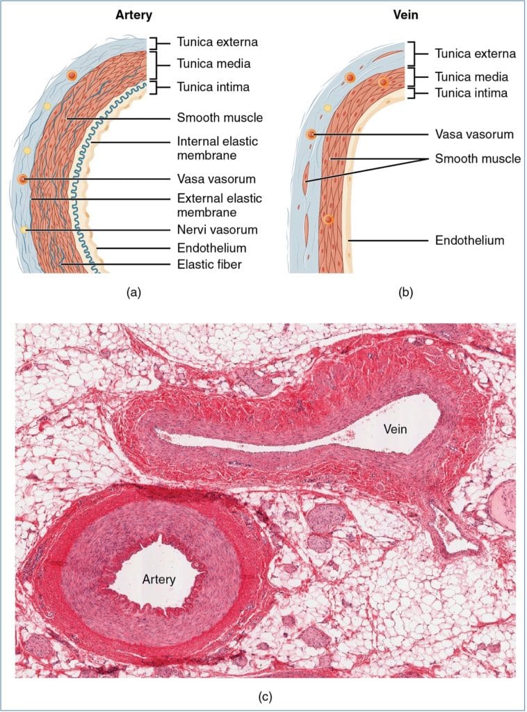
Vessel Wall Structure
Tunica Intima
- Innermost layer in direct contact with blood
- Composed of endothelium (simple squamous epithelium)
- Larger vessels also contain a sub-endothelial layer
Tunica Media
- Thickest layer: smooth muscle + elastin
- Circular smooth muscle regulates tone
- Controlled by sympathetic nervous system
- Responsible for vasoconstriction and vasodilation
Tunica Externa (Adventitia)
- Outermost collagen-rich layer
- Contains:
- Nerve fibres
- Lymphatics
- Vasa vasorum (in larger vessels)
Flow, Pressure & Vessel Dynamics
Relationships
- Vessel diameter affects cross-sectional area, local pressure, and flow velocity
- As total cross-sectional area increases (e.g. in capillaries), velocity decreases
- Smaller vessels offer higher resistance and modulate blood distribution
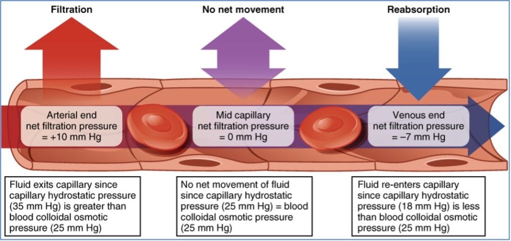
Fluid Exchange Across Capillaries
Key Forces Governing Fluid Movement
- Capillary Hydrostatic Pressure
- Drives fluid out of capillaries
- ~35 mmHg (arterial end) → ~15 mmHg (venous end)
- Net hydrostatic pressure = capillary hydrostatic pressure – interstitial pressure (~0 mmHg)
- Colloid Osmotic Pressure
- Draws fluid into capillaries via plasma proteins
- ~25 mmHg (constant along vessel length)
- Net osmotic pressure = capillary osmotic pressure – interstitial osmotic pressure (~1 mmHg)
Net Filtration Pressure = Net Hydrostatic Pressure – Net Osmotic Pressure
- Fluid leaves at arterial end, reabsorbed at venous end
Oedema
Definition
- Excess interstitial fluid accumulation → visible swelling
Causes
- ↑ Fluid exit from capillaries
- ↓ Fluid reabsorption into blood
Mechanisms
- ↓ Plasma proteins → reduced oncotic pull
- ↑ Capillary hydrostatic pressure:
- Incompetent valves
- Local obstruction
- Congestive heart failure (e.g. pulmonary oedema)
- High blood volume
- ↑ Capillary permeability:
- Due to inflammation
Vascular Injury and Disease
Atherosclerosis
- Formation of fatty plaques in the subendothelial space
- May ulcerate, trigger thrombosis, and obstruct flow

Aneurysms
- Loss of elasticity in vessel wall
- Leads to bulging and potential rupture
- Common in elastic arteries

Arterial Dissections
- Blood enters between vessel wall layers
- Can occlude the lumen and cause ischemia

Summary – Blood Vessel Physiology
Blood vessel physiology explains how vessel structure and function regulate flow, pressure, and fluid exchange across the vascular system. From resistance modulation in arterioles to reabsorption at capillary beds, these dynamics are vital for systemic equilibrium. For a broader context, see our Cardiovascular Overview page.