Table of Contents
Overview – Heart Anatomy
Heart anatomy refers to the structural organization of the heart within the thoracic cavity, including its chambers, layers, valves, blood vessels, and coverings. Understanding this anatomy is essential for interpreting cardiovascular physiology, diagnosing structural abnormalities, and performing clinical procedures like auscultation or ECG placement. This article provides a high-yield, clinically relevant overview tailored for final-year medical students.
Definition
The heart is a muscular organ responsible for pumping blood through the pulmonary and systemic circulations. It lies within the middle mediastinum of the thorax and is encased in a fibrous sac known as the pericardium.
Anatomical Location
- Located in the middle mediastinum, within the inferior mediastinum
- Extends from the 2nd rib to the 5th intercostal space
- Anterior to vertebral column, posterior to sternum
- Flanked by both lungs and rests on the diaphragm
- Two-thirds of the heart lies to the left of the midsternal line
- The middle mediastinum also contains the pericardium, great vessels, trachea, and esophagus


Pericardium (Coverings of the Heart)
- Double-walled sac enclosing the heart
- Contains serous fluid for lubrication
- Fibrous Pericardium:
- Tough connective tissue
- Anchors and protects the heart
- Prevents overfilling (e.g. in cardiac tamponade)
- Serous Pericardium:
- Parietal layer: lines fibrous pericardium
- Visceral layer (epicardium): adheres to external heart surface
Layers of the Heart Wall
- Epicardium: outermost layer; visceral pericardium
- Myocardium: muscular layer; responsible for contraction
- Endocardium: endothelial lining of chambers; prevents clotting and acts as a barrier between blood and myocardium

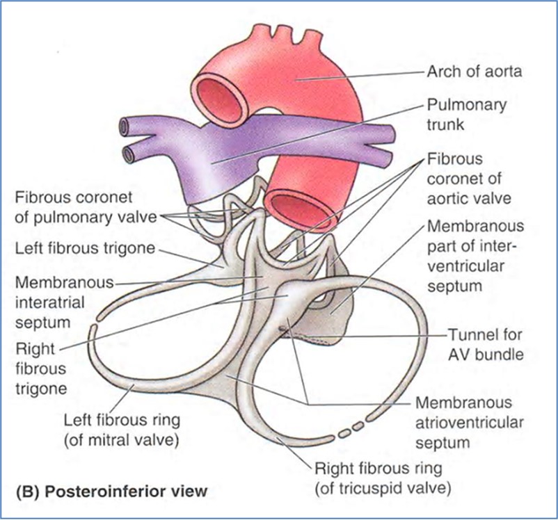
Fibrous Skeleton of the Heart
- Dense connective tissue network of collagen and elastin
- Functions:
- Anchors muscle fibers, valves, and great vessels
- Reinforces myocardium
- Electrically isolates atria from ventricles
Components:
- Septums: separate chambers (interatrial, atrioventricular, interventricular); non-conductive
- Rings: surround valves and great vessels to prevent dilation under pressure
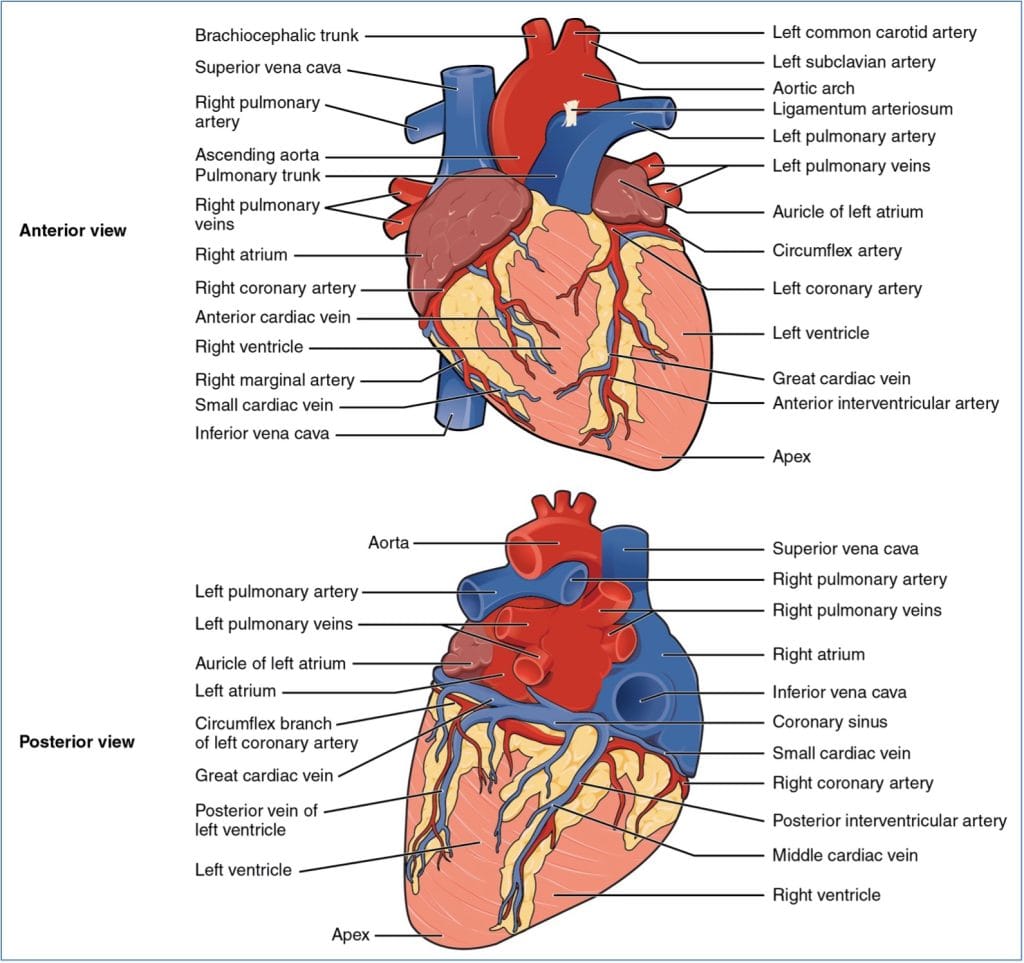
Chambers & Great Vessels
Atria (Superior Chambers)
- Thin-walled receiving chambers
- Auricles: increase atrial volume
- Right Atrium:
- Receives blood from:
- Superior vena cava
- Inferior vena cava
- Coronary sinus
- Features: pectinate muscles
- Receives blood from:
- Left Atrium:
- Receives oxygenated blood from 4 pulmonary veins
Ventricles (Inferior Chambers)
- Thick-walled discharging chambers
- Trabeculae carneae line internal surfaces
- Papillary muscles attach to valves
- Right Ventricle:
- Forms most of anterior surface
- Pumps to lungs via pulmonary trunk
- Left Ventricle:
- Forms postero-inferior surface
- Pumps to body via aorta

Heart Landmarks
- Coronary Sulcus (AV groove): encircles heart; houses coronary arteries and veins
- Anterior Interventricular Sulcus: contains LAD; separates ventricles anteriorly
- Posterior Interventricular Sulcus: contains PDA; separates ventricles posteriorly
Pathway of Blood Through the Heart
- Right heart → pulmonary circuit (deoxygenated blood to lungs)
- Left heart → systemic circuit (oxygenated blood to body)
- Colour change reflects gas exchange:
- Pulmonary: blue to red (O₂ gain)
- Systemic: red to blue (O₂ loss)

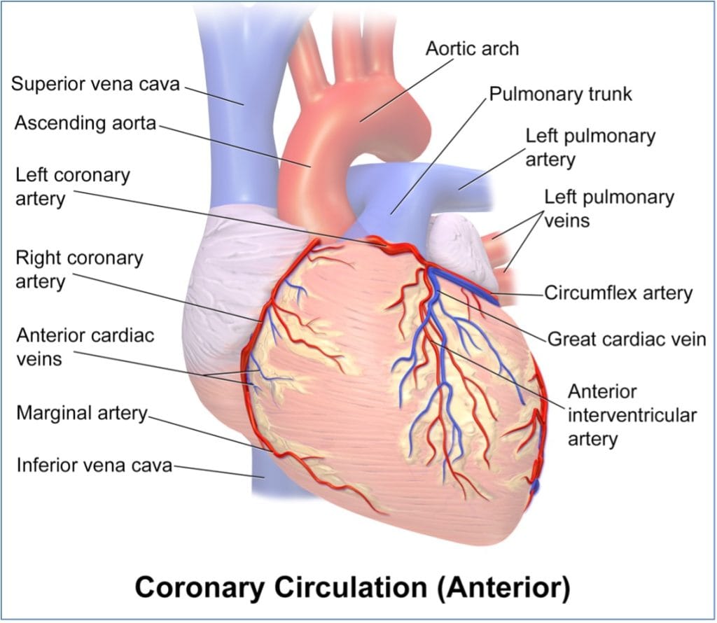
Coronary Circulation
Arterial Supply
- Coronary arteries arise from aorta and run in epicardium
- Left Coronary Artery:
- LAD (Anterior Interventricular Artery): apex, anterior LV, 2/3 IV septum
- Circumflex Artery: left atrium, lateral LV
- Right Coronary Artery:
- Marginal Artery: right lateral myocardium
- Posterior Interventricular Artery: posterior ventricular walls
- Forms anastomosis with LAD
Venous Drainage
- Collected by Great, Middle, and Small Cardiac Veins
- Drain into coronary sinus, then right atrium
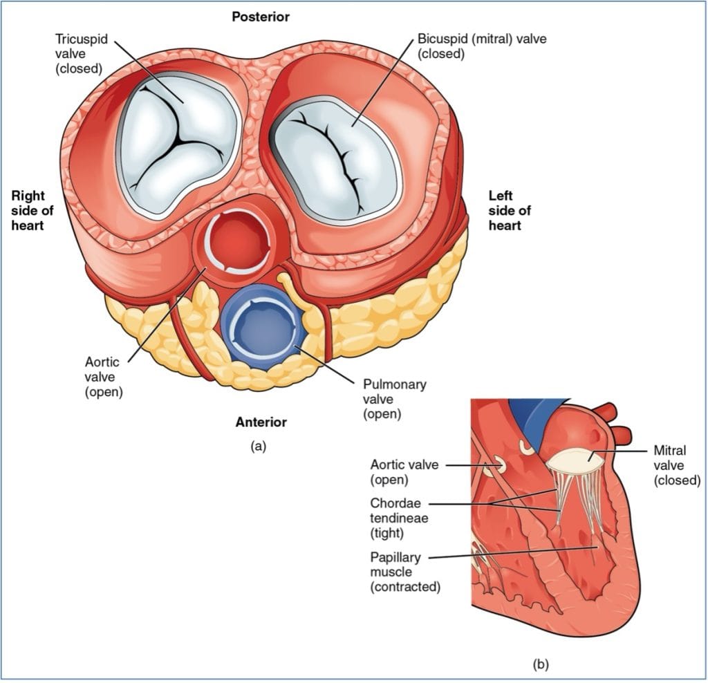
Heart Valves
Atrioventricular (AV) Valves
- Located between atria and ventricles
- Prevent backflow during ventricular contraction
- Chordae tendineae anchor cusps to papillary muscles
- Tricuspid Valve: right side; 3 cusps
- Mitral Valve: left side; 2 cusps
Semilunar (SL) Valves
- Located at outflow of ventricles
- Open under ventricular pressure
- Pulmonary Valve: RV → Pulmonary trunk
- Aortic Valve: LV → Aorta
Valve Positions During Ventricular Contraction
Valve Positions During Ventricular Relaxation

Valve Sounds
- “Lubb” (S1): closure of AV valves (M1 + T1)
- “Dupp” (S2): closure of SL valves (A2 + P2)
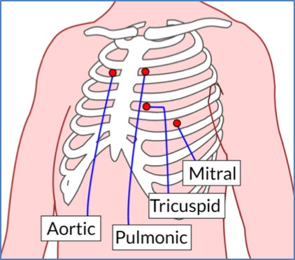
Auscultation Landmarks
- Aortic, Pulmonary, Tricuspid, and Mitral valve sounds can be heard at distinct points on the thorax based on flow direction and valve position
Where to Listen to Heart Sounds
Summary – Heart Anatomy
Heart anatomy encompasses the detailed structure of the heart, including its chambers, valves, coverings, and vascular supply, all essential for maintaining efficient circulation and interpreting clinical findings. A foundational understanding of heart anatomy supports diagnostics and skills such as auscultation, ECG interpretation, and cardiovascular imaging. For a broader context, see our Cardiovascular Overview page.