Table of Contents
Overview – Thoracic Anatomy
Thoracic anatomy forms the foundation for understanding the respiratory and cardiovascular systems. The thorax provides mechanical protection for vital organs, structural support for respiration, and passageways for major vessels and nerves. Thoracic anatomy is clinically important because injuries or pathology within this region can compromise breathing, circulation, or nervous system function.
Thoracic Overview
- 3 Main Parts:
- Thoracic Cage – skeletal framework
- Thoracic Wall – muscular components
- Thoracic Cavity – internal area containing organs
- 3 Internal Compartments:
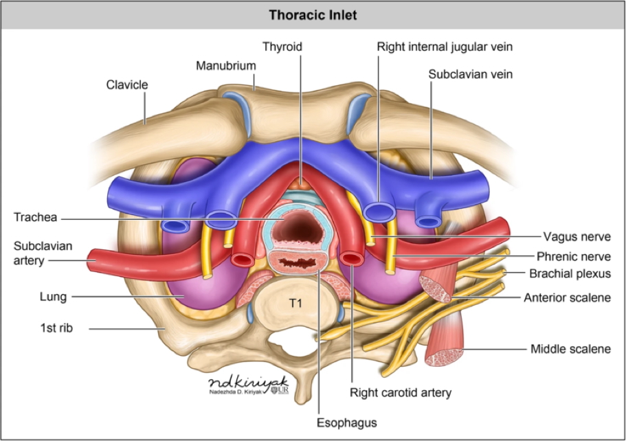
- Mediastinum (central compartment): heart, oesophagus, trachea, major vessels, nerves
- Left Pleural Cavity: left lung
- Right Pleural Cavity: right lung


Relationship of Thorax to Other Regions
- Neck connections: trachea, oesophagus, major vessels and nerves
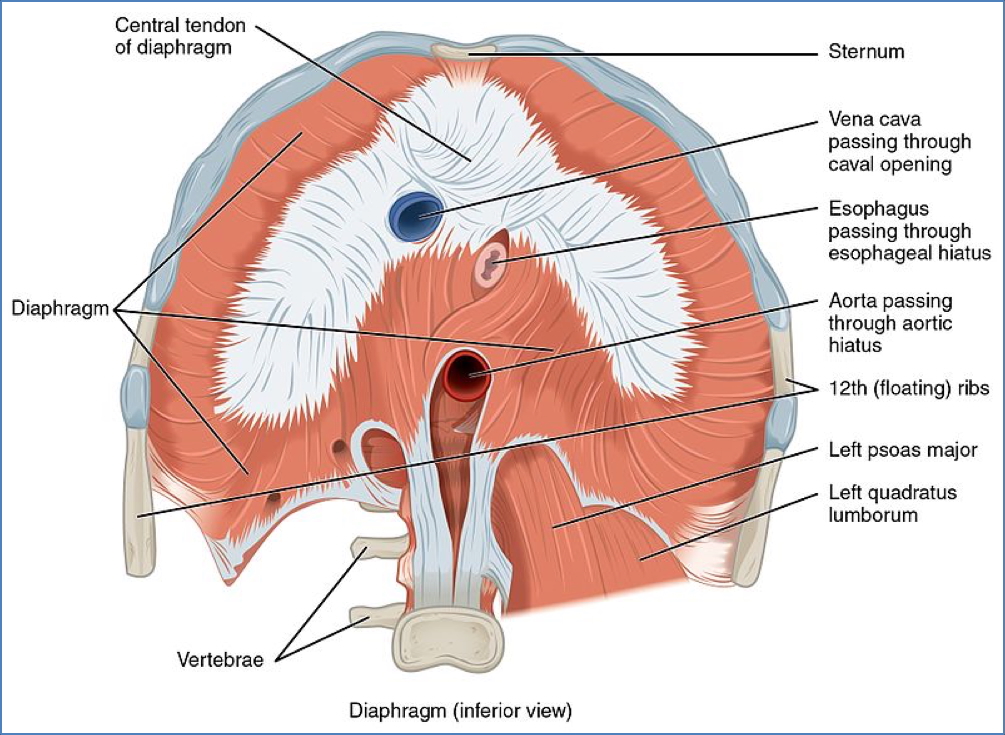
- Abdominal connections: oesophagus, inferior vena cava, aorta
Thoracic Skeleton
Vertebrae
- 12 thoracic vertebrae (T1–T12)
- Distinguishing features:
- Heart-shaped vertebral body (extra weight-bearing)
- Inferiorly projecting spinous process (permits rib mobility)
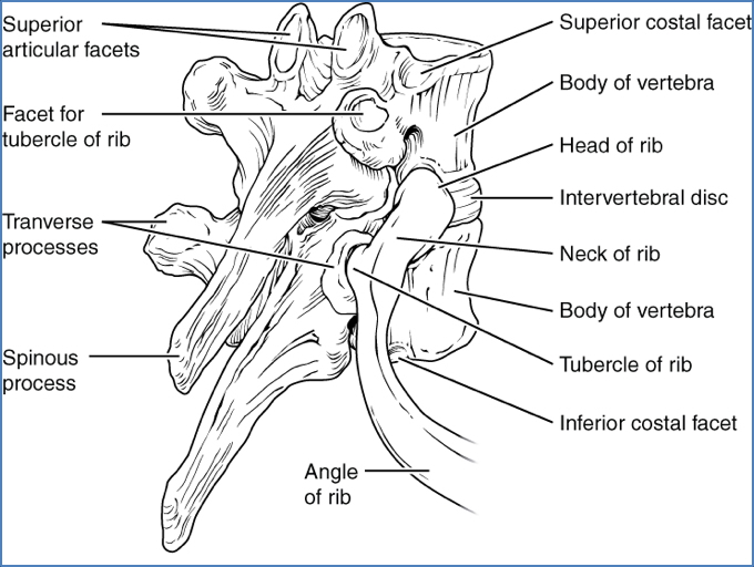
- Large transverse processes (rib articulation)
- Costal demifacets for rib heads


Sternum
- Three parts: manubrium, body, xiphoid process
- Sternal angle (angle of Louis): junction of manubrium and body
- Landmark for: tracheal bifurcation, aortic arch, T4 vertebra, 2nd rib
- Articulations:
- Ribs 1–7: sternocostal (synovial) joints
- Ribs 8–10: interchondral joints (indirect attachment)
Note: Thoracic cage joints allow small movements; combined, these create mobility for breathing.

Ribs
- 12 pairs of ribs:
- Ribs 1–7 = True ribs (direct sternum attachment)
- Ribs 8–12 = False ribs (indirect or no sternum attachment)
- Ribs 11–12 = Floating ribs (insert into abdominal muscles)
- Features:
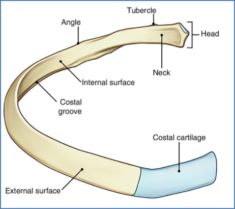
- Posterior end: head, neck, tubercle (for vertebral articulation)
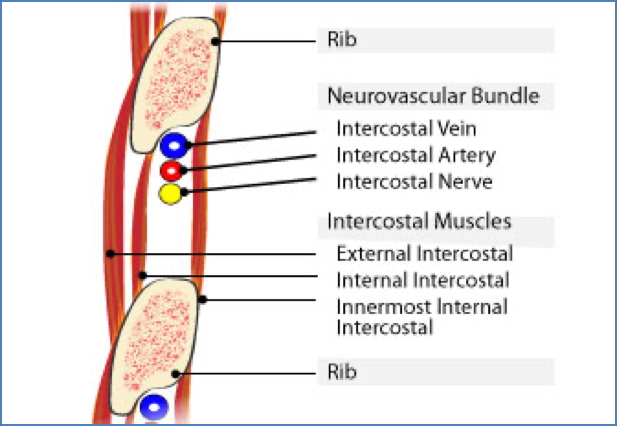
- Shaft: thin, flat, vertically oriented with subcostal groove (houses intercostal nerve, artery, vein)
- Anterior end: attaches via costal cartilage
- Typical articulations:
- Rib head with its own vertebra and the vertebra above
- Tubercle with transverse process of corresponding vertebra

Atypical Ribs
- Rib 1: horizontal, short, articulates only with T1, scalene tubercle, grooves for subclavian vessels
- Rib 2: horizontal, otherwise typical
- Rib 10: articulates only with its own vertebra (single facet)
- Ribs 11 & 12: articulate only with their own vertebra, no tubercles or anterior attachment

Muscular Components
The thoracic wall consists of three layers of intercostal muscles:
- External intercostal muscles
- Oriented infero-anteriorly (diagonal)
- Incomplete anteriorly → transition to anterior intercostal membrane
- Internal intercostal muscles
- Deep to externals
- Transition posteriorly into the posterior intercostal membrane
- Innermost intercostal muscles
- Oriented infero-posteriorly
- Incomplete posteriorly

Blood Supply
- Posterior intercostal arteries (branches of descending aorta)
- Anterior intercostal arteries (branches of internal thoracic arteries from subclavian)
Nerve Supply
- Anterior rami of thoracic spinal nerves supply the intercostal muscles segmentally.
Accessory Muscles of Respiration
- Inspiration:
- Scalene muscles
- Sternocleidomastoid
- External intercostals
- Mechanism: elevate ribs and sternum → pump-handle and bucket-handle movements
- Expiration:
- Abdominal wall muscles (↑ intra-abdominal pressure → diaphragm pushed up)
- Internal intercostals (depress ribs and sternum → reverse pump/bucket-handle)
The Diaphragm
The diaphragm is the primary muscle of respiration.
- Structure: musculotendinous sheet dividing thorax and abdomen
- Action:
- Contraction → flattens downward → inspiration
- Relaxation → domes upward → expiration
- Origins: xiphoid process, costal margin (7th rib), lower ribs 11–12, body of T12
- Insertion: central tendon
- Blood supply: superior & inferior phrenic arteries
- Venous drainage: brachiocephalic veins, azygous veins, inferior vena cava
- Nerve supply: phrenic nerve (C3–C5)
- Receives sympathetic fibres from cervical ganglia
- Provides both voluntary and autonomic control


Pleura
- Continuous serous sacs: visceral pleura (covers lungs) & parietal pleura (lines thoracic cavity)
- Between layers: pleural space with lubricating serous fluid
- Surface tension keeps lungs inflated and pleurae together


- Costodiaphragmatic recess: potential space for lung expansion during forced inspiration

Thoracic Movements of Breathing
- Breathing movements arise from combined actions of diaphragm, thoracic wall, and accessory muscles.
- Upper 6 ribs: pump-handle action → increases anteroposterior diameter
- Lower 6 ribs: bucket-handle action → increases transverse diameter

Summary – Thoracic Anatomy
Thoracic anatomy integrates the skeletal cage, muscles, and cavity housing critical organs like the lungs and heart. Understanding rib articulations, pleural spaces, and diaphragm mechanics is vital for respiratory medicine. Explore more high-yield anatomy content on our Respiratory Overview page.