Table of Contents
Overview – The Skeleton
The skeleton provides the structural framework for the body and is essential for support, movement, protection of vital organs, and mineral and fat storage. It also plays a central role in blood cell formation through bone marrow. Understanding the structure, classification, and organisation of the skeleton is foundational knowledge for medical students studying the musculoskeletal system.
Functions
- Support – Framework for muscles and organs.
- Protection – Shields vital organs (e.g. brain, heart, lungs).
- Movement – Facilitates motion through articulation and muscle attachment.
- Storage:
- Minerals – Especially calcium (Ca²⁺), vital for metabolic processes and tightly regulated in the bloodstream.
- Marrow – Stores both haematopoietic stem cells (for blood production) and fat.
- Blood Production – Haematopoiesis in red bone marrow.
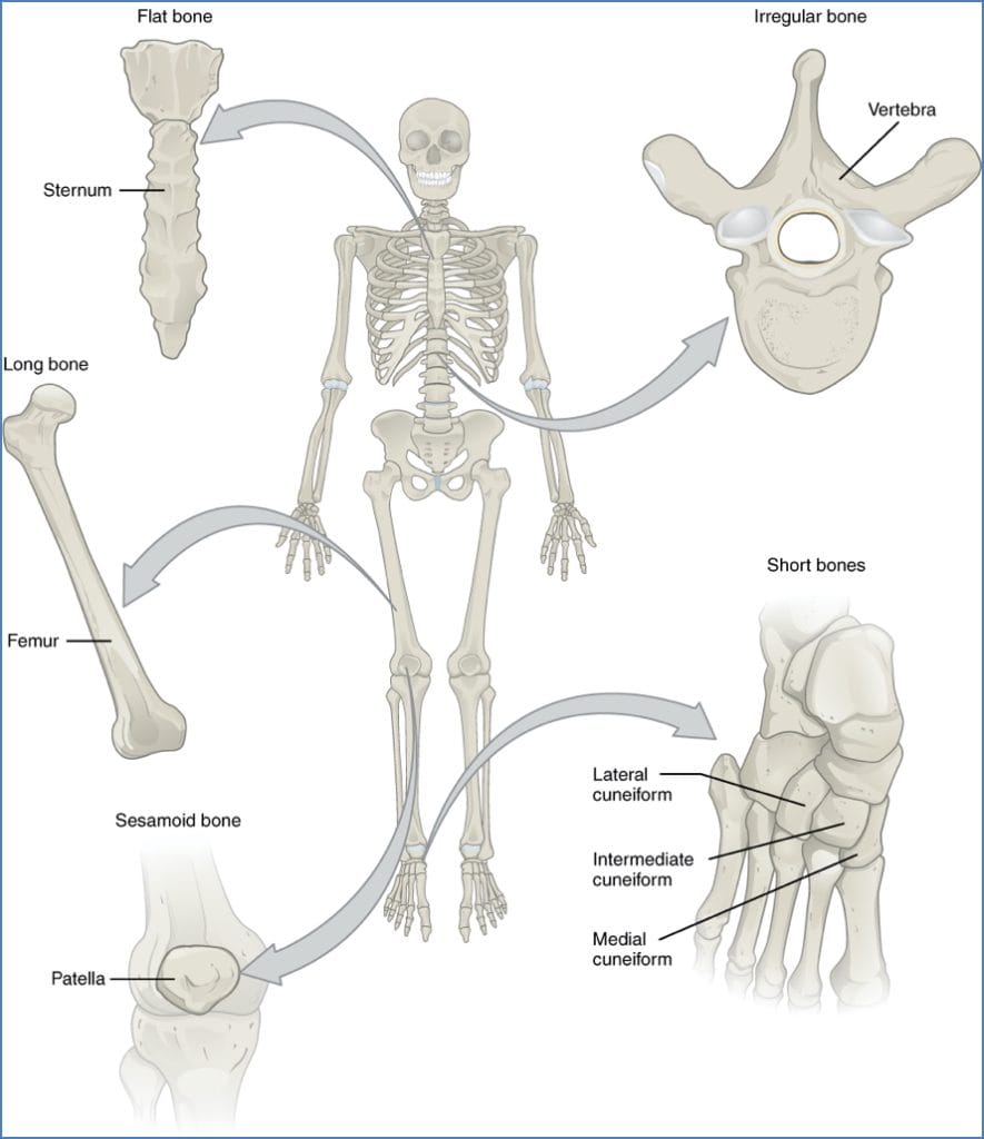
Classification of Bones
Bones are classified based on shape and function:
- Long Bones – Length > width; function in leverage and movement.
- Examples: Humerus, femur
- Short Bones – Nearly equal in length and width; stability with limited motion.
- Examples: Carpals, tarsals
- Flat Bones – Thin, often curved; provide protection and surface area for muscle attachment.
- Examples: Sternum, scapula
- Irregular Bones – Complex shapes; serve various protective and structural functions.
- Examples: Vertebrae, innominate bones (ossa coxae)
- Sesamoid Bones – Embedded within tendons; protect tendons and increase mechanical advantage.
- Examples: Patella, occasional sesamoids in the tendons of the big toe
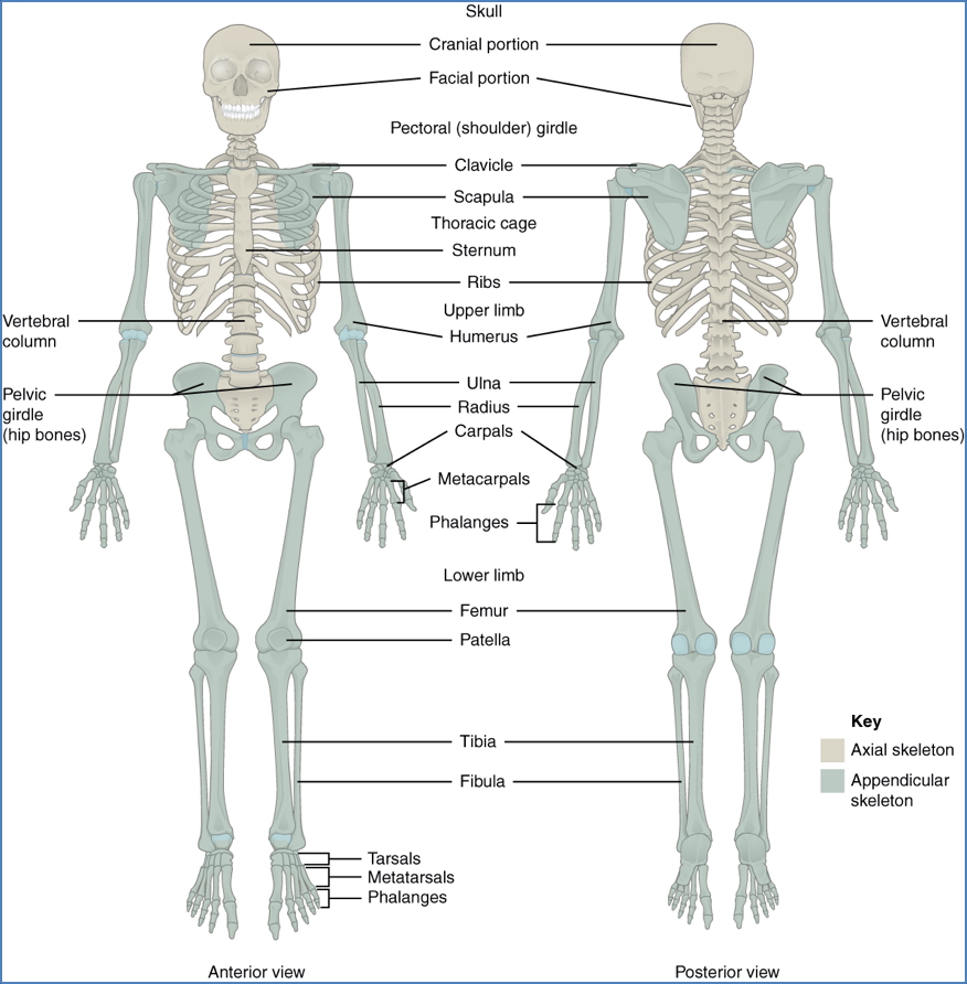
Axial vs Appendicular Skeleton
- Axial Skeleton:
- Forms the central axis.
- Includes: Skull, vertebral column, ribs, sternum.
- Appendicular Skeleton:
- Includes limbs and girdles.
- Upper and lower limbs + pectoral and pelvic girdles.
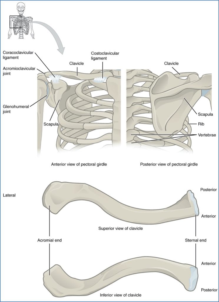
Pectoral Girdle
- Composed of the clavicle and scapula.
- Connects upper limbs to the axial skeleton.
- Provides mobility and structural support.
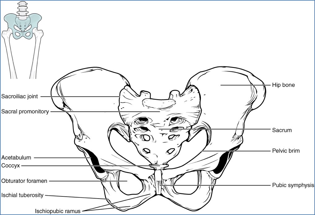
Pelvic Girdle
- Formed by two coxal (hip) bones, one on each side of the sacrum.
- Each coxal bone consists of three fused components:
- Ilium
- Ischium
- Pubis
- Connected anteriorly by the pubic symphysis.
- Also referred to as:
- Ossa coxae
- Innominate bones
- Function: Anchors lower limbs to the axial skeleton and protects pelvic organs.
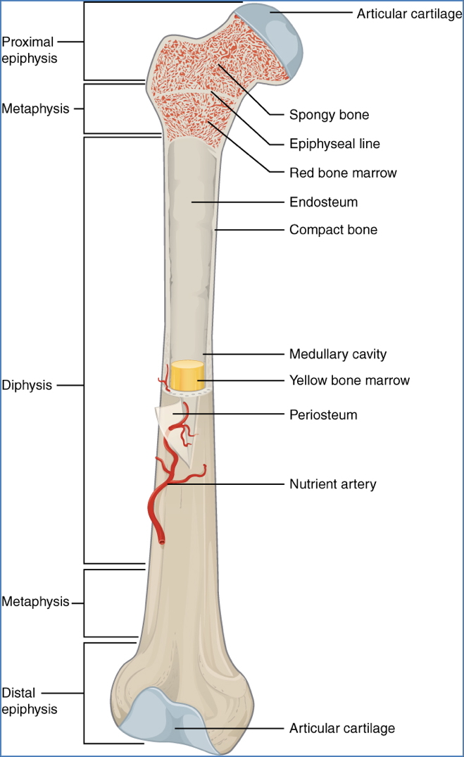
Long Bone Structure
- Diaphysis
- Shaft of the bone.
- Hollow → provides strength while minimising weight.
- Contains yellow marrow (fat storage).
- Epiphysis
- Expanded proximal and distal ends of long bones.
- Covered in articular cartilage.
- Boundary marked by the epiphyseal line (growth plate remnant).
- Medullary (Marrow) Cavity
- Central cavity of the diaphysis.
- Contains red marrow (for haematopoiesis) and yellow marrow.
- Lightens the bone while retaining strength.
- Periosteum
- Outer fibrous connective tissue layer.
- Contains Sharpey’s fibres for anchoring.
- Inner cellular layer contains:
- Osteoblasts (bone formation)
- Osteoclasts (bone resorption)
- Outer fibrous connective tissue layer.
- Nutrient Foramen
- External opening for blood vessels and nerves.
- More numerous near the epiphyseal region.
- Essential for bone vascular supply.

Summary – The Skeleton
The skeleton is a structural and functional foundation of the human body, supporting movement, protecting organs, storing minerals, and producing blood cells. It is organised into the axial and appendicular skeletons and made up of bones classified by shape and function. Mastering skeletal anatomy is critical for any musculoskeletal clinical understanding. For a broader context, see our Musculoskeletal Overview page.