Table of Contents
Overview – Vitiligo
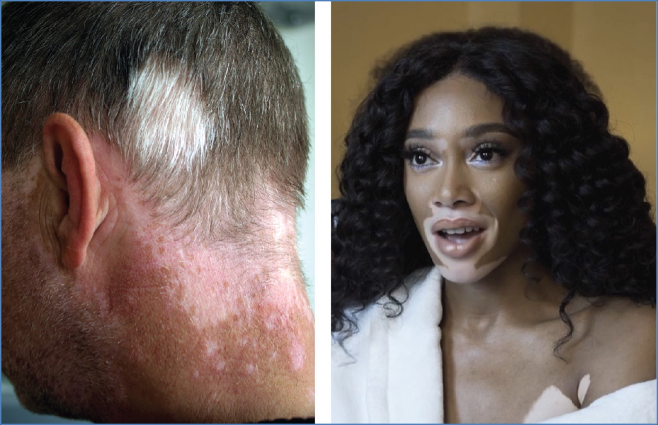
Vitiligo is a chronic, acquired pigmentary disorder characterised by the progressive loss of melanocytes, resulting in well-defined, milky-white patches of depigmented skin. While it is not physically harmful, it can have significant psychosocial effects, particularly in darker skin types. It is commonly associated with other autoimmune diseases, such as thyroid disorders and type 1 diabetes. Understanding vitiligo is crucial for final-year medical students, particularly in dermatology, general medicine, and primary care, where the condition often presents first.
Definition
Vitiligo is a chronic depigmenting disorder caused by the loss of functional melanocytes in the epidermis, resulting in patches of depigmented (leukodermic) skin.
Aetiology
- Systemic autoimmune disorder
- Involves autoantibodies targeting melanocytes
- Often associated with the enzyme tyrosinase (TYR), which is key in melanin synthesis
- Strong links with other autoimmune conditions
Epidemiology
- Affects 0.5–1% of the global population
- Can begin at any age, but >50% of cases start before the age of 20
- No sex or ethnic predilection, though more cosmetically apparent in darker skin tones
Pathophysiology
- Autoimmune destruction of melanocytes in the epidermis
- The tyrosinase enzyme (TYR), involved in melanin synthesis, becomes a target
- T-cell mediated immune response → melanocyte apoptosis
- Loss of pigment in affected skin areas
- Genetic susceptibility with associated HLA markers
- Often co-exists with other autoimmune conditions
Clinical Features
- Depigmented macules/patches:
- Well-demarcated, milky-white
- Symmetrical or segmental
- Common sites:
- Periorificial areas (mouth, eyes, genitals)
- Bony prominences (hands, elbows, knees)
- Progressive course:
- Gradual enlargement of lesions
- New lesions may develop after skin trauma (Koebner phenomenon)
- Associated conditions:
- Autoimmune thyroid disease (most common)
- Type 1 diabetes mellitus
- Pernicious anaemia
- Addison’s disease
- Systemic lupus erythematosus (SLE)
- Psoriasis
- Alopecia areata
Investigations
- Clinical diagnosis (mainly visual)
- Wood’s lamp: Enhances contrast of depigmented lesions
- Autoimmune screen:
- Thyroid function tests (TFTs)
- Blood glucose, HbA1c
- Vitamin B12, ANA if autoimmune suspicion
- Skin biopsy (rarely needed):
- Absence of melanocytes in epidermis
Management
- General measures:
- Minimise trauma to skin
- Cosmetic camouflage (e.g. makeup, micropigmentation)
- Sun protection (to avoid sunburn of depigmented areas)
- Topical therapy:
- Potent corticosteroids (first-line)
- Calcineurin inhibitors (e.g. tacrolimus for facial lesions)
- Phototherapy:
- Narrowband UVB is the mainstay for widespread or rapidly progressing vitiligo
- Systemic therapy (in extensive or refractory cases):
- Oral steroids
- Immunomodulators (e.g. methotrexate, ciclosporin)
- Minocycline (inhibits oxidative stress)
- Targeted therapy:
- Ruxolitinib (JAK inhibitor) — approved for facial vitiligo
- Depigmentation therapy:
- For extensive vitiligo in dark-skinned patients
- E.g. monobenzyl ether of hydroquinone
Complications
- Psychosocial distress and reduced quality of life
- Increased risk of sunburn in depigmented areas
- Co-existing autoimmune conditions
- Stigma and social withdrawal
Differential Diagnosis
- Pityriasis alba (especially in children)
- Post-inflammatory hypopigmentation
- Tinea versicolor
- Chemical leucoderma
- Morphea
- Albinism (generalised pigment loss from birth)
Summary – Vitiligo
Vitiligo is an autoimmune condition leading to destruction of melanocytes and resultant well-demarcated white patches of skin. It may be associated with systemic autoimmune diseases and can significantly impact quality of life. Early recognition, appropriate investigations, and a tailored management approach are crucial. For a broader context, see our Skin & Dermatology Overview page.