Table of Contents
Overview – Testicular Tumours
Testicular tumours are the most common malignancy in young adult males, especially between the ages of 15 and 40. Although rare overall, they are clinically significant due to their potential for early metastasis and impact on fertility. Most cases are germ cell tumours, which are further classified into seminomas and non-seminomatous germ cell tumours. Recognising the typical presentation—painless unilateral testicular mass—is critical for early detection and successful treatment.
Definition
Testicular tumours are abnormal proliferations of cells arising within the testis. They are most commonly derived from germ cells and can behave in either a benign or malignant fashion. The majority are malignant, and most originate from germinal epithelium.
Aetiology
- Idiopathic in most cases
- Cryptorchidism (undescended testes)
- In utero oestrogen exposure
- Genetic predisposition
- Environmental and hormonal influences
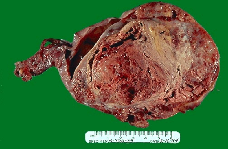
Morphology / Pathophysiology
- Originates from germ cells
- Classified into:
- Seminomas (uniform cell population)
- Non-seminomatous germ cell tumours (NSGCTs) (heterogeneous histology – may include embryonal carcinoma, yolk sac tumour, choriocarcinoma, or teratocarcinoma)
- Spread is typically via lymphatics to the retroperitoneal lymph nodes
- Hormonal activity (e.g. hCG production) can cause systemic effects such as gynaecomastia
Clinical Features
Symptoms
- Painless, unilateral testicular mass
- May be associated with hydrocele
- In some cases, hormonal effects such as breast tenderness or gynaecomastia
Complications
- Retroperitoneal mass from lymph node metastasis
- Infertility
- Systemic metastases if diagnosis delayed
Types of Germ Cell Tumours
Seminoma
- ~40% of testicular tumours
- Most common in males aged 30–50 years
- Slow-growing but malignant
- No tumour markers (AFP or hCG usually normal)
- Treatment: Orchidectomy ± radiotherapy or chemotherapy
- Prognosis:
- Excellent response to treatment
- Cure rate >90%
Non-Seminomatous Germ Cell Tumours (NSGCTs)
Embryonal Carcinoma
- ~25% of testicular tumours, common in children under 4 years
- Highly malignant with early metastasis
- Tumour markers: Elevated alpha-fetoprotein (AFP) and human chorionic gonadotropin (hCG)
- Treatment: Orchidectomy + combination chemotherapy
- Prognosis: Poorer than seminomas due to aggressive nature
Teratocarcinoma
- Contains multiple tissue types (ectoderm, mesoderm, endoderm)
- Often part of a mixed germ cell tumour
- Behaviour depends on degree of differentiation
Investigations
- Scrotal ultrasound – first-line imaging
- Tumour markers:
- AFP (raised in NSGCTs)
- hCG (may be raised in both seminomas and NSGCTs)
- LDH (non-specific marker of tumour burden)
- CT chest/abdomen/pelvis – to assess for metastasis
- Histological diagnosis via orchidectomy specimen
Management
- Radical inguinal orchidectomy (diagnostic and therapeutic)
- Seminomas: Radiation and/or chemotherapy
- NSGCTs: Chemotherapy (cisplatin-based regimens)
- Regular tumour marker monitoring for recurrence
- Fertility preservation should be discussed prior to treatment
Complications
- Metastasis (lungs, liver, retroperitoneum)
- Infertility due to tumour or treatment
- Hormonal effects (e.g. gynaecomastia from hCG-secreting tumours)
- Psychological impact of cancer diagnosis and orchiectomy
Differential Diagnosis
- Epididymal cyst
- Hydrocele
- Varicocele
- Epididymo-orchitis
- Testicular torsion
Summary – Testicular Tumours
Testicular tumours are important malignancies to recognise early, particularly in young males. Testicular tumours typically present as painless testicular enlargement and are categorised as seminomas or non-seminomatous germ cell tumours. While seminomas have excellent treatment outcomes, non-seminomatous variants are more aggressive. For a broader context, see our Reproductive Health Overview page.