Table of Contents
Overview – Muscle Physiology
Muscle physiology is the study of how muscles contract, generate force, and respond to nervous stimulation. It covers key processes such as excitation-contraction coupling, the sliding filament mechanism, and the function of motor units. Mastery of these mechanisms is essential for clinical understanding of muscular diseases, neuromuscular junction disorders, and physical performance.
Events at the Neuromuscular Junction
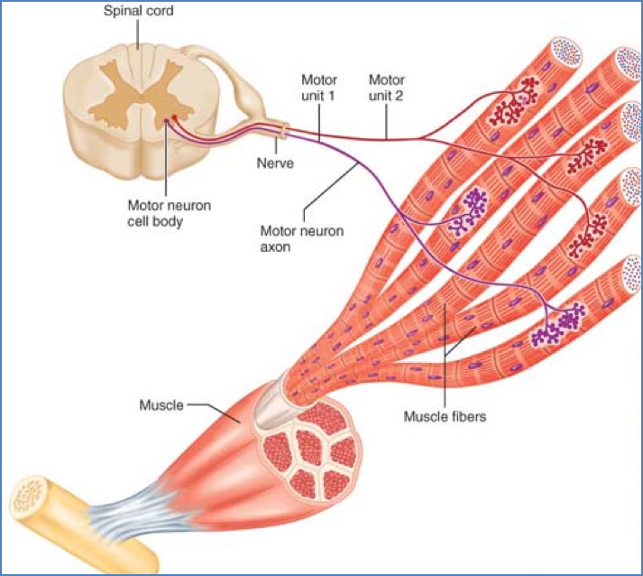
- Each somatic motor neuron branches to innervate multiple muscle fibres.
- Each muscle fibre has one neuromuscular junction, typically midway along its length.
Steps:
- Nerve impulse reaches axon terminal
- Voltage-gated Ca²⁺ channels open → Ca²⁺ enters terminal
- Acetylcholine (ACh) vesicles released into synaptic cleft
- ACh binds sarcolemma receptors → action potential initiated
- ACh is rapidly degraded by acetylcholinesterase
- Action potential spreads along sarcolemma → into T-tubules
- Triggers Ca²⁺ release from terminal cisternae of sarcoplasmic reticulum (SR)
- Ca²⁺ binds to troponin, displacing tropomyosin from actin binding sites
- Myosin heads bind actin → contraction begins
- Ca²⁺ is actively reabsorbed by SR
- Troponin-tropomyosin complex re-forms → contraction ends

Sliding Filament Theory
Resting State
- Low intracellular Ca²⁺
- Tropomyosin blocks myosin-binding sites on actin
- Muscle is relaxed
Contraction Cycle (in presence of Ca²⁺ and ATP):
- Excitation–Cross Bridge Formation
- Ca²⁺ binds troponin → exposes actin-binding sites
- Myosin head binds actin → actomyosin complex forms
- Power Stroke
- Pi release → myosin head pivots (70°), pulling actin
- Muscle shortens, tension develops
- Cross-Bridge Detachment
- New ATP binds → myosin releases actin
- Re-Cocking of Myosin Head
- ATP hydrolysed → myosin returns to high-energy position
- Ready for next contraction cycle


Relaxation
- Nerve signal stops
- Ca²⁺ reabsorbed by SR (ATP-dependent pump)
- Binding sites covered → muscle returns to resting state
The Motor Unit
- A motor unit = one motor neuron + all muscle fibres it innervates
- Fine control (e.g. fingers, eyes): Small motor units
- Gross movement (e.g. thighs): Large motor units
- Allows graded contraction by varying recruitment
Muscle Twitch
- A motor unit’s response to a single action potential
- 3 Phases:
- Latent Period – Excitation-contraction coupling begins
- Contraction – Cross-bridge cycling; tension rises (10–100 ms)
- Relaxation – Ca²⁺ reuptake; tension falls

Graded Muscle Responses
1. Stimulation Frequency
- Rapid successive stimuli → summation of twitches
- Leads to smooth sustained contraction

2. Motor Unit Recruitment
- Stronger stimulus → recruits more motor units
- Greater fibre activation → stronger contraction


Muscle Tone
- Sustained, low-level contraction in resting muscles
- Maintains posture, joint stability, and readiness
- Mediated by reflexes responding to stretch receptors
Determinants of Contractile Force
1. Number of Fibres Stimulated
- More motor units → greater force
2. Size of Fibres
- Larger fibres (greater cross-sectional area) generate more force
- Muscle hypertrophy = cellular enlargement
3. Frequency of Stimulation
- Increased frequency → Ca²⁺ accumulation → stronger contractions
- Maximum = tetanus
4. Muscle Length (Stretch)
- Optimal force at resting length where all myosin heads can bind actin

5. Muscle Fibre Type
- White fibres:
- Anaerobic
- Large CSA
- High force
- Red fibres:
- Aerobic
- Mitochondria-rich
- Lower force but sustained endurance
Summary – Muscle Physiology
Muscle physiology explains how skeletal muscles convert neural signals into forceful movement. This involves excitation-contraction coupling at the neuromuscular junction, the sliding filament model of contraction, and the dynamic function of motor units. Understanding these principles is critical for interpreting both normal movement and neuromuscular dysfunction. For a broader context, see our Musculoskeletal Overview page.