Table of Contents
Overview – Genetics of Development
Genetics of development refers to the molecular mechanisms that drive the transformation of a single fertilised egg into a highly complex, multicellular human organism. This topic is central to embryology, anatomy, and clinical genetics, explaining how genes guide processes such as cell differentiation, organ formation, and body patterning. Understanding the genetic control of development is crucial in identifying the origins of congenital anomalies, syndromes, and genetic diseases.
Definition
Genetic control of development encompasses the genetically driven processes that govern cellular behaviour, tissue formation, and organogenesis during embryonic development. It involves tightly regulated gene expression, signalling pathways, transcription factors, and cellular interactions.
Key Developmental Processes
Cellular Processes
- Cell proliferation → Rapid mitotic division to expand cell numbers.
- Cell differentiation → Specialisation of cells via changes in gene expression.
- Cell–cell interactions → Communication between cells to coordinate responses.
- Cell migration → Rearrangement into functional tissues and organ systems.
Cell Fate & Differentiation
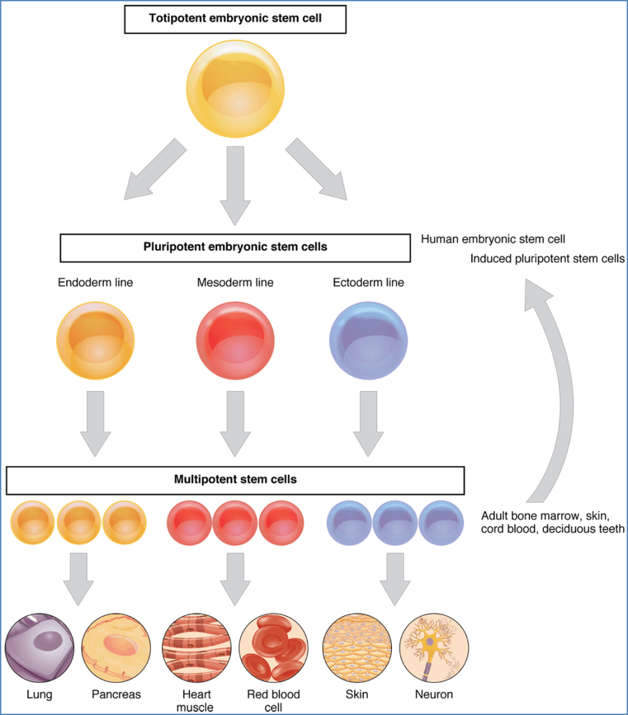
From Totipotency to Specialisation
- Totipotent cells → Can become any human cell (early embryogenesis).
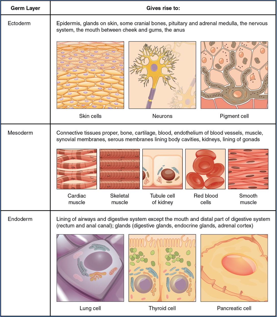
- Pluripotent cells (germ layers) → Have committed pathways but are not yet specialised.
- Terminally differentiated cells → Fully specialised with restricted function.
2. CNX OpenStax, CC BY 4.0 <https://creativecommons.org/licenses/by/4.0>, via Wikimedia Commons
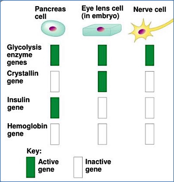
Cell Lineage
- Lineage determines future specialisation.
- All embryonic cells express core survival genes.
- Only a subset of genes is active in any given cell type (e.g., insulin gene in pancreas cells).
Morphogens & Spatial Information
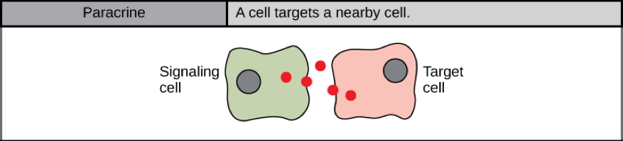
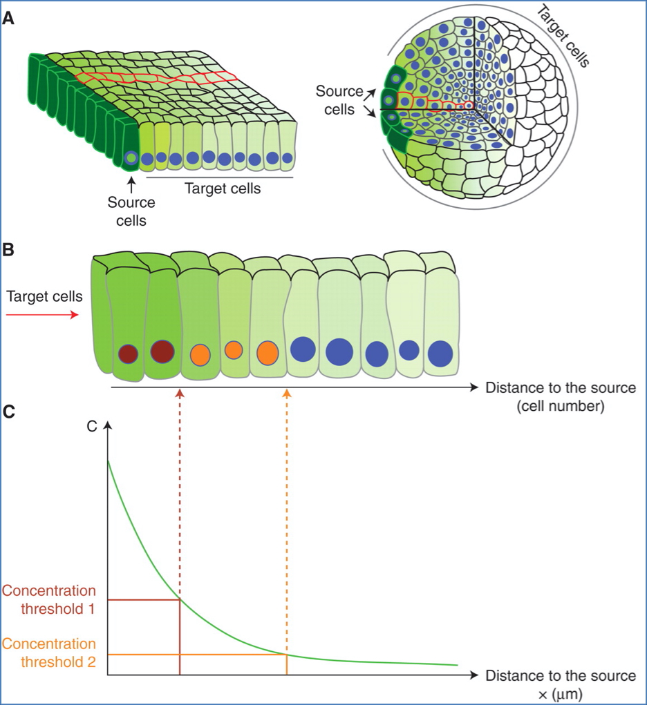
What Are Morphogens?
- Secreted proteins that act on nearby cells (paracrine signalling).
- Determine cell fate by:
- Concentration thresholds.
- Combinatorial exposure from different organiser cells.
Transmission Modes
- Diffusible molecules → Bind receptors on nearby cells.
- Extracellular matrix signalling → Ligand travels via the matrix.
- Direct contact → Gap junction communication between cells.
Molecular Basis of Differentiation
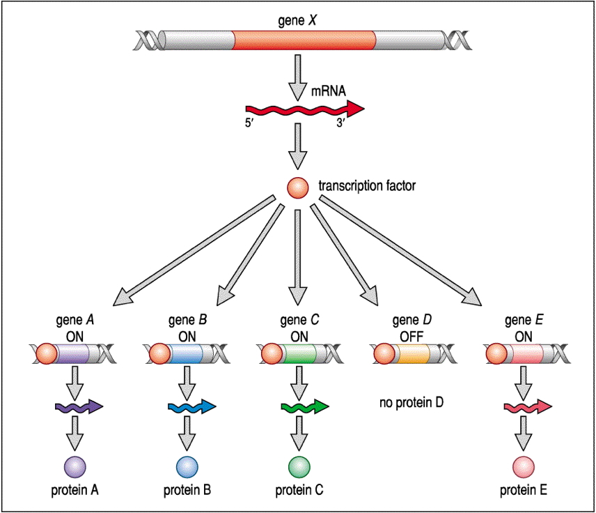
Gene Expression Control
- Signals such as hormones, positional cues, or morphogens activate transcription factors.
- Transcription factors control tissue-specific gene sets.
- Histone modification:
- Tags open or close parts of the genome.
- Epigenetic memory is passed on during mitosis → ensures heritable function.

Inductive Signalling in Development
Key Regulatory Concepts
- Cellular induction → Reactant cell is prompted to differentiate.
- Time restriction → Signal only effective during a specific developmental window.
- Space restriction → Induction requires proximity.
- Reciprocal induction → Reactant and inductor cells mutually influence each other.
- Sequential induction → Cascade where newly differentiated cells induce others.
Body Plan Formation
What is the “Body Plan”?
- The body plan is the early developmental layout of an organism.
- In vertebrates, this includes segmentation into repeating units (somites), which later give rise to specific anatomical structures.
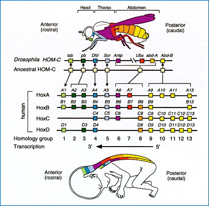
Role of HOX Genes
- Homeobox (HOX) genes direct the body plan layout by controlling regional identity along the anterior-posterior axis.
- Humans have 39 HOX genes, functioning as transcription factors.
- HOX proteins regulate expression of tissue-specific genes → drive cellular differentiation into muscles, bones, etc.
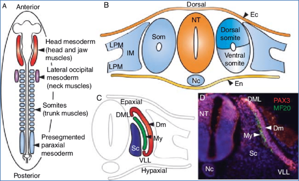
Segmentation and Somite Development
- Early human embryos are segmented like other vertebrates.
- Somites (blocks of mesoderm):
- Differentiate into vertebrae, ribs, and back muscles.
- Each segment’s identity is defined by a specific combination of HOX gene expression.
Mechanism of HOX Gene Action
- HOX gene → HOX protein (transcription factor) → binds DNA → activates/inhibits downstream genes.
- Result: Cell populations differentiate appropriately depending on the spatial and temporal HOX expression.

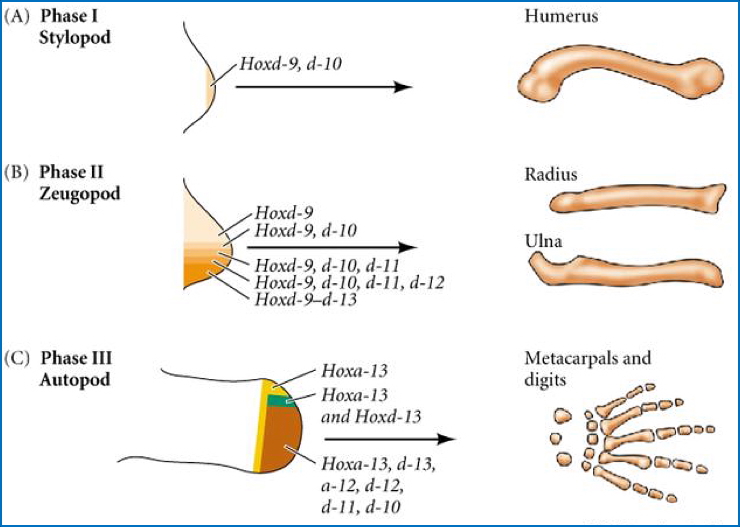
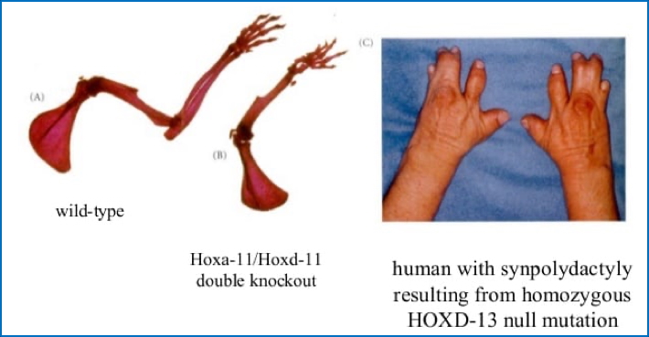
Example: HOX D Genes in Limb Development
- HOXD gene cluster governs upper limb patterning:
- HOXD9 & HOXD10: expressed early and persist.
- HOXD13: expressed later, guiding digit formation.
- Different HOX genes turn on at different times and for different durations, allowing for spatial patterning.

Developmental Abnormalities from HOX D Malexpression
- Knockout or mutation of a HOX D gene → loss of the limb part it controls.
- Partial errors → malformations like syndactyly (fused digits) or synpolydactyly (fused + extra digits).
Organogenesis and Master Control Genes
What is Organogenesis?
- The process by which different cell types organize into fully functional organs.
- Requires tight genetic regulation for coordinated development.
Master Control Genes
- These genes trigger gene cascades directing organ-specific proliferation and differentiation.
- A single master gene can regulate dozens of downstream targets.
Example: PAX6 and Eye Development
- PAX6 is the master control gene for eye and neural tube development.
- Highly conserved across species (e.g., humans, mice, flies).
- Mutations in PAX6:
- In humans → aniridia (absence of the iris) due to haploinsufficiency (50% loss of function).
- In mice → complete loss of both alleles → no eyes.
- Inserting PAX6 in tissue can even produce ectopic eye structures.

Sex Determination and the SRY Gene
The Genetic Basis of Sex
- Chromosomal sex:
- Male = XY
- Female = XX
- Phenotypic sex is determined by presence or absence of the SRY gene on the Y chromosome.
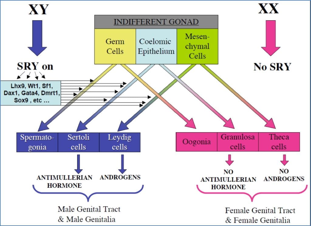
Role of SRY
- Encodes testis-determining factor (TDF), a transcription factor.
- If present → undifferentiated gonads become testes → secrete testosterone → development of male genitalia.
- If absent → default pathway is female.
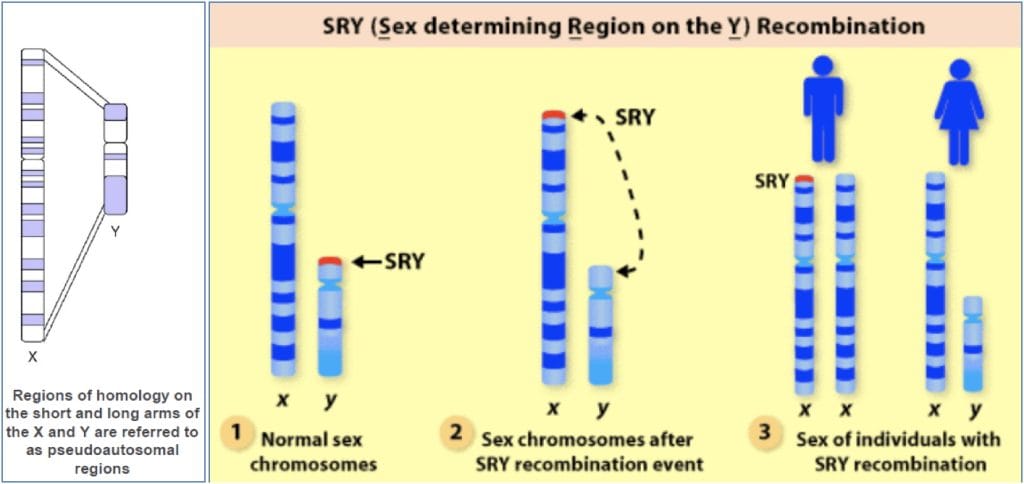
Genetic Anomalies
- XY females or XX males may result from translocation of the SRY gene between sex chromosomes.
- SRY is located near the pseudoautosomal region of the Y chromosome, making translocations more likely.
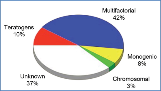
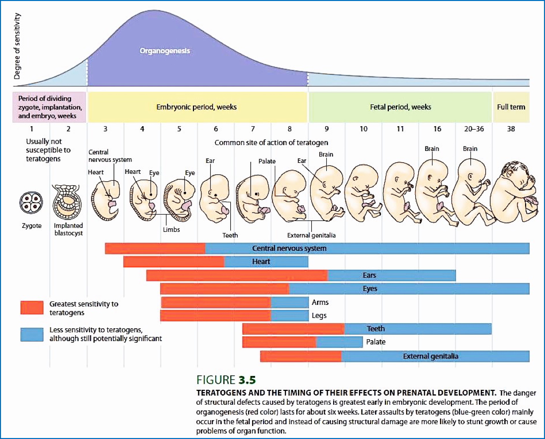
Teratogens and Birth Defects
What Are Teratogens?
- Teratogens are environmental agents that disrupt normal embryonic development.
- Effects depend on timing:
- Organogenesis is the most vulnerable period due to rapid cell division.
- Different organs have specific windows of susceptibility.
Impact of Teratogens
- Can cause structural anomalies, functional deficits, or spontaneous miscarriage.
- Common examples: medications, radiation, infections, and maternal metabolic conditions.
Summary – Genetics of Development
The genetic control of development underpins all stages of embryogenesis, from totipotent stem cells to complex organs. Key players include transcription factors, morphogens, HOX genes, and master control genes like Pax6, all of which guide spatial, temporal, and functional development. Errors in these processes can result in congenital anomalies or disrupted body plans. For a broader context, see our Genetics & Cancer Overview page.