Table of Contents
Overview – Sialolithiasis
Sialolithiasis refers to the formation of calculi (stones) within the ducts or parenchyma of the salivary glands. It most commonly affects the submandibular gland and presents with pain and swelling that typically worsens during meals due to stimulated salivation. Though often self-limiting, persistent cases may require surgical intervention, especially if glandular involvement is present.
Definition
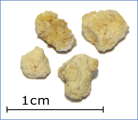
- Ductal stone composed primarily of hydroxyapatite within a salivary gland.
- Leads to obstruction and chronic sialadenitis.
- Distribution:
- 80% in the submandibular gland
- <20% in the parotid gland
- ~1% in the sublingual gland
Risk Factors
- Dehydration
- Diabetes mellitus
- Alcohol use
- Medications causing dry mouth (e.g. anticholinergics)
Clinical Features
- Pain and tenderness over the affected gland
- Swelling following meals due to stimulated salivation
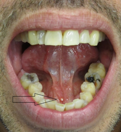
- Palpable calculus on intraoral examination (often floor of mouth for submandibular stones)
Investigations
- Sialogram – outlines the ductal system and may show obstruction
- CT scan – confirms stone presence and location, especially useful for non-palpable or intraglandular stones
Management
- Conservative:
- Increase hydration and salivation (e.g. sour candies)
- Gentle gland massage
- Interventional:
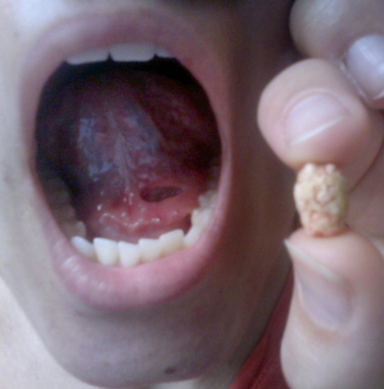
- Ductal dilation and stone removal via transoral excision
- If stone is located within the gland → surgical excision of the entire gland
Summary – Sialolithiasis
Sialolithiasis involves the obstruction of salivary flow due to stone formation, most often in the submandibular gland. It presents with meal-related glandular swelling and pain. Diagnosis is clinical, supported by imaging, and treatment ranges from conservative approaches to surgical excision in more persistent cases.
For broader context, visit our Head and Neck Overview page, or return to the Gastrointestinal Overview.