Table of Contents
Overview – Neuronal Physiology
Neuronal physiology underpins all nervous system communication. Neurons transmit signals through rapid electrical impulses and specialised chemical synapses, enabling responses in other neurons, muscle, or glandular tissue. This article covers the mechanics of action potentials, synaptic transmission, and post-synaptic receptor types — foundational concepts in neurology, pharmacology, and clinical neuroscience.
What is Neurotransmission?
- Definition: Communication between a neuron and another cell (neuron, muscle, organ, etc.)
- Occurs at: The synapse
- Synapse Components:
- Pre-synaptic neuron = sends signal
- Synaptic cleft = gap between neurons
- Post-synaptic cell = receives signal
- Synaptic potential = depolarising trigger for neurotransmitter release
Synapse Types
Neurons can synapse in different anatomical locations for varying effects:
- Axo-dendritic: Axon → dendrite (most common; integrates multiple inputs)
- Axo-somatic: Axon → soma (modulatory influence)
- Axo-axonic: Axon → axon (controls neurotransmitter release)

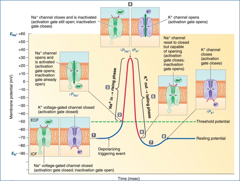
Action Potential: Ionic Basis
Key Ions:
- Na⁺ (influx): Initiates depolarisation
- K⁺ (efflux): Restores resting potential (repolarisation + hyperpolarisation)
- Ca²⁺ (influx): Triggers vesicle exocytosis at synapse
Phases of Action Potential:
- Resting State: All gated channels closed
- Threshold: Na⁺ channels open → depolarisation
- Peak: Na⁺ channels inactivate, K⁺ channels open
- Repolarisation: K⁺ exits → returns toward baseline
- Hyperpolarisation: Continued K⁺ efflux
- Resetting: K⁺ channels close; Na⁺ channels reset
Refractory Periods
- Absolute: No new AP possible (Na⁺ inactivated)
- Relative: New AP possible only with larger stimulus
Conduction Speed – What Affects It?
- Axon diameter: Larger = faster conduction
- Myelination: Saltatory conduction at nodes of Ranvier accelerates signal transmission
Phases of Synaptic Transmission
- Arrival of AP → voltage-gated Ca²⁺ channels open at axon terminal
- Ca²⁺ influx → triggers neurotransmitter vesicle exocytosis
- Neurotransmitter release (ACh, glutamate, GABA, etc.)
- NT diffuses across cleft and binds post-synaptic receptors
- Graded potential generated:
- Excitatory (EPSP): Depolarisation
- Inhibitory (IPSP): Hyperpolarisation
- Signal summation: If threshold met at axon hillock → new AP
- Termination: NT degraded (e.g. AChE) or reabsorbed into pre-synaptic neuron


Types of Post-Synaptic Receptors
1. Ionotropic Receptors (Ligand-Gated Ion Channels)
- Fast, direct action
- Binding of NT → opens ion channel → alters membrane potential
- Excitatory: Na⁺ / Ca²⁺ influx → EPSP
- Inhibitory: Cl⁻ influx or K⁺ efflux → IPSP
2. Metabotropic Receptors (G-Protein Coupled Receptors)
- Slower, indirect
- NT binding → G-protein activation → 2nd messengers (e.g. cAMP)
- Alters ion channels, gene expression, metabolism
- Used in modulatory pathways (e.g. dopamine, serotonin)

Functional Outcomes of Neurotransmission
- Direct Response:
- NMJ → muscle contraction
- Sympathetic input → ↑ heart rate
- Sensory Relay:
- Peripheral neuron → spinal cord → thalamus → cortex
- Modulation:
- One neuron alters strength/speed of another’s signal
Summary – Neuronal Physiology
Neuronal physiology is driven by precise control of ion flux, membrane potentials, and neurotransmitter signalling. Action potentials and synapses allow rapid, targeted communication across the nervous system. The diversity of receptor types and synapse configurations enables both fast responses and complex regulation. For related anatomical organisation, see our Nervous System page.