Table of Contents
Overview – Nervous System Organization
This nervous system overview describes the nervous system as the master control and communication system of the human body. It integrates sensory input, coordinates organ function, and orchestrates voluntary and involuntary responses to internal and external stimuli. Structurally, it comprises the brain, spinal cord, peripheral nerves, and sensory organs. Functionally, it can be divided into sensory (afferent) and motor (efferent) pathways, with further distinction between somatic and autonomic subsystems. Understanding this high-level organisation provides a critical foundation for studying neurology, neuroanatomy, and neurophysiology in clinical contexts.
Macrostructures of the Nervous System
- Brain
- Spinal Cord
- Peripheral Nerves
- Special Sense Organs:
- Eyes – vision
- Ears – hearing & balance
- Tongue – taste
- Olfactory bulbs – smell
- Skin – touch, pressure, pain, temperature
Core Functions
- Detection of internal and external stimuli
- Transmission and integration of information
- Coordination of responses in muscles, glands, and organs
- Maintenance of homeostasis via rapid signalling
Organisation of the Nervous System
Central Nervous System (CNS)
“The CPU & motherboard”
- Brain
- Spinal cord
- Integration centre for sensory data and motor commands
Peripheral Nervous System (PNS)
“The cables”
- Cranial nerves and spinal nerves
- Communication lines between CNS and the rest of the body
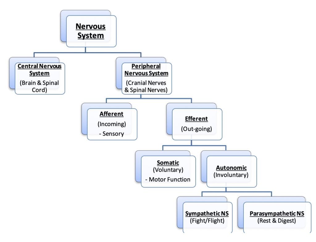
Functional Divisions of the PNS
- Afferent Division (Sensory)
- Carries incoming information from sensory receptors to the CNS
- Efferent Division (Motor)
- Carries outgoing commands from the CNS to effectors (muscles & glands)
Efferent Subdivisions:
- Somatic Nervous System
- Voluntary motor control (e.g., skeletal muscles)
- Autonomic Nervous System
- Involuntary control (e.g., heart, glands, smooth muscle)
- Sympathetic Division: “Fight or flight”
- Parasympathetic Division: “Rest and digest”
Refer to the flowchart above for visual hierarchy.
The Neuron – Structural Overview
- Dendrites (Receptive field): Receive input from other neurons
- Soma (Cell body): Processes graded potentials
- Axon (Efferent projection): Conducts action potentials
- May be myelinated or unmyelinated
- Axon hillock: Trigger zone for action potential initiation
- Myelin sheath: Insulation for faster conduction
- Nodes of Ranvier: Gaps between myelin sheaths allowing saltatory conduction
- Synaptic terminals: Release neurotransmitters to target tissues

Neuroglia – Nervous System Support Cells
Non-neuronal cells that support, protect, and maintain homeostasis
Central Nervous System Glia:
- Astrocytes:
- Nutrient transport between neurons & capillaries
- Guide migrating neurons during development
- Synapse formation, ionic and neurotransmitter buffering
- Microglia:
- Immune surveillance
- Respond to injury or infection by phagocytosis
- Oligodendrocytes:
- Form myelin sheaths in CNS
- Ependymal Cells:
- Line CNS ventricles and spinal canal
- Circulate cerebrospinal fluid (CSF) with cilia
Peripheral Nervous System Glia:
- Schwann Cells:
- Myelinate axons in the PNS
- Aid axonal regeneration after injury
- Satellite Cells:
- Surround neuron cell bodies in ganglia
- Provide structural and metabolic support

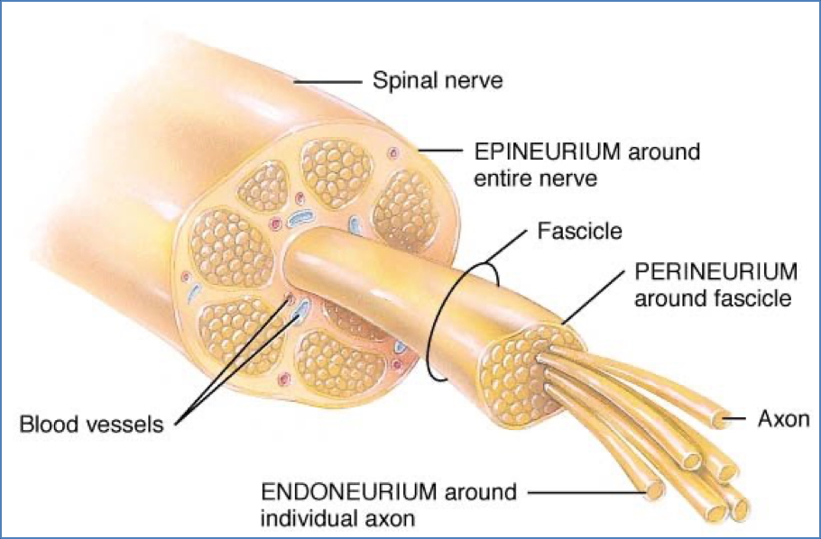
Connective Tissue Layers of Peripheral Nerves
- Endoneurium:
- Delicate tissue surrounding individual axons
- Perineurium:
- Bundles axons into fascicles
- Epineurium:
- Tough outer sheath enclosing multiple fascicles
- Contains blood vessels
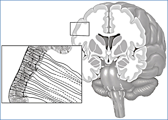
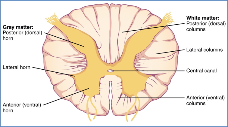
Gray Matter vs White Matter
Gray Matter
- Composed of neuron cell bodies (soma) and neuroglia
- Located in:
- Cerebral cortex
- Spinal cord centre (H-shaped zone)
- Ganglia / Nuclei
White Matter
- Comprised of myelinated axons
- White due to lipid-rich myelin
- Found in:
- Peripheral nerves & plexuses
- Central tracts of brain and spinal cord
Ganglia
- Definition: Clusters of neuron cell bodies in the PNS
- Types:
- Dorsal root ganglia (Afferent): Contain sensory neuron bodies
- Sympathetic ganglia (Efferent): Contain autonomic motor neuron bodies
- CNS Equivalents: Called nuclei or basal nuclei
- Essential for reflex arcs, motor control, and autonomic signalling

Spinal Nerves & Dermatomes
- Each spinal nerve provides motor and sensory innervation to a segment of the body.
- Dermatome: The area of skin innervated by sensory fibres of a single spinal nerve
- Maps provide clinical reference for spinal nerve injury or pathology
- Arise from segmentation of mesoderm during embryonic development


Summary – Nervous System Overview
The nervous system integrates and processes stimuli from both the external and internal environments through a complex but highly structured network. Divided into central and peripheral components, and further into afferent and efferent divisions, it enables everything from conscious movement to autonomic function. Neurons and supporting glial cells operate in synchrony, protected and organised by connective tissue and segmented anatomical pathways. For a broader context, see our Nervous System page.