Table of Contents
Overview – Motor Pathways
Motor pathways are the descending neural tracts responsible for transmitting movement commands from the brain to skeletal muscles. They are divided into lateral and ventromedial systems, controlling voluntary and involuntary/postural movements respectively. Understanding their anatomy, decussation points, and target neurons is crucial for clinical neurology and lesion localisation.
Lateral Motor Pathways
Function: Voluntary movement of distal extremities (hands and feet)
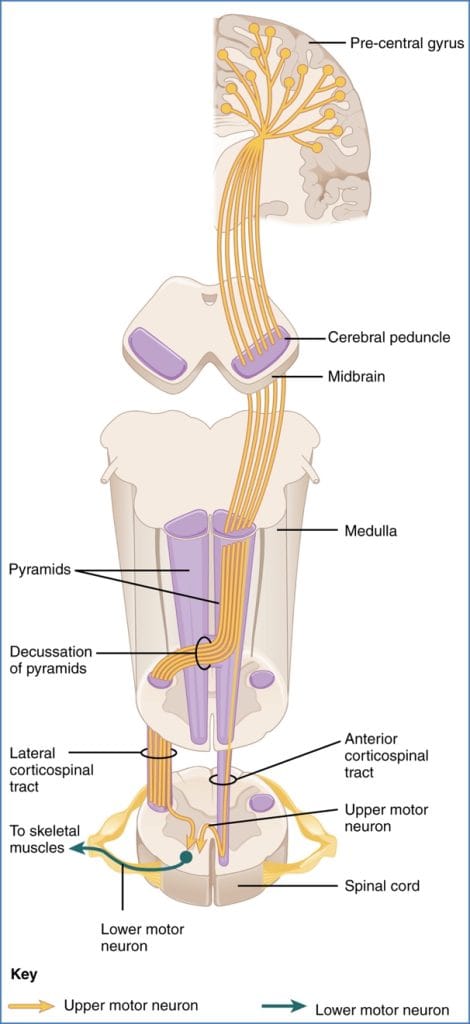
1. Corticospinal Tract
- Origin: Primary motor cortex (M1)
- Course:
- Passes through internal capsule
- Continues through brainstem
- Decussates at the medullary pyramids (pyramidal decussation)
- Terminates: Ventral horn of spinal cord
- Control: Fine voluntary movement of distal limbs
2. Rubrospinal Tract
- Origin: Red nucleus (midbrain)
- Decussation: Immediately after red nucleus (pons level)
- Path: Travels down lateral white matter of spinal cord
- Terminates: Ventral horn (spinal grey matter)
- Function: Facilitates distal motor control, especially in upper limbs
More prominent in animals; humans rely primarily on corticospinal tract
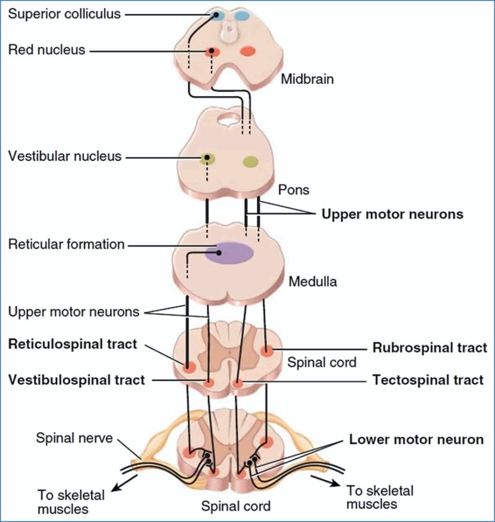
Ventromedial (Extrapyramidal) Pathways
Function: Involuntary control of posture, balance, and large trunk/proximal muscles
1. Tectospinal Tract
- Origin: Superior colliculus (midbrain)
- Decussation: Between midbrain and pons
- Function: Coordinates visual tracking, reflexive head/eye movements
2. Vestibulospinal Tract
- Origin: Vestibular nuclei (medulla)
- Decussation: None (remains ipsilateral)
- Function: Maintains balance and upright posture
3. Reticulospinal Tracts (Pontine & Medullary)
- Origin: Pontine reticular formation
- Decussation: None
- Function: Regulate muscle tone, autonomic posture control, visceral motor function

Termination & Integration
- Upper motor neurons from descending tracts terminate in the ventral horn of spinal grey matter
- They may synapse with:
- Spinal interneurons:
- May be part of central pattern generators (e.g. rhythmic walking)
- Lower motor neurons (LMNs):
- Project directly to skeletal muscles via peripheral nerves
- Spinal interneurons:
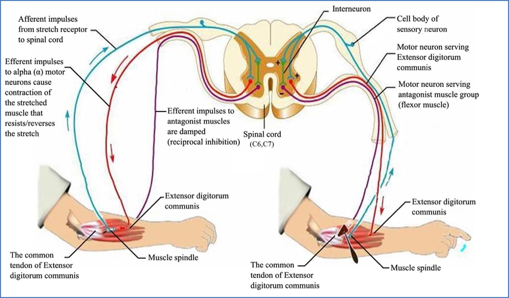
Reflexes
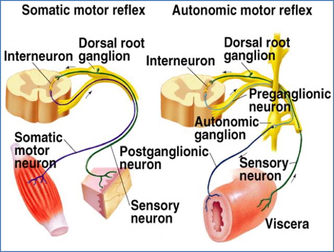
Somatic Reflexes
- Rapid, involuntary motor responses to sensory stimuli
- Occur via a reflex arc:
- Receptor
- Sensory neuron
- Integration centre (spinal cord or brainstem)
- Motor neuron
- Effector (skeletal muscle)
Visceral Reflexes (Autonomic)
- Regulate internal organ function (e.g. BP, gut motility)
- Components:
- Visceral sensory neuron (e.g. stretch, pH, chemical irritation)
- Ganglionic neuron (integration)
- Motor neuron
- Effector (smooth muscle, cardiac muscle, glands)

Summary – Motor Pathways
Motor pathways consist of descending tracts that control voluntary and involuntary movements. Lateral pathways like the corticospinal tract are essential for fine motor control, while ventromedial tracts manage posture and reflexive movement. Upper motor neurons synapse with spinal interneurons or lower motor neurons, integrating motor commands and reflexes. For a broader context, see our Nervous System Overview page.