Table of Contents
Overview – Hearing and Equilibrium
The ear plays a dual sensory role: detecting sound (hearing) and maintaining balance (equilibrium). These functions are mediated by specialised mechanoreceptors called hair cells, located within the cochlear and vestibular apparatus of the inner ear. Hearing involves transmission and transduction of sound waves, while equilibrium monitors the position and movement of the head to coordinate balance and posture. Disorders of either system can result in deafness, vertigo, or gait disturbance—making mastery of this anatomy and physiology essential in clinical neurology and ENT.
Ear Anatomy
Outer Ear (Air-Filled)
- Pinna (Auricle): Funnels sound into external auditory canal
- External Auditory Canal: Conducts sound to tympanic membrane
- Contains ceruminous glands → secretes cerumen (earwax)
- Tympanic Membrane: Vibrates in response to sound
Middle Ear (Air-Filled)
- Auditory Ossicles:
- Malleus → Incus → Stapes
- Amplify sound ≈20x before reaching inner ear
- Stapedius & Tensor Tympani Muscles: Reflexively dampen loud sounds
- Oval Window: Receives vibration from stapes → cochlea
- Eustachian Tube: Equalises pressure between middle ear and nasopharynx

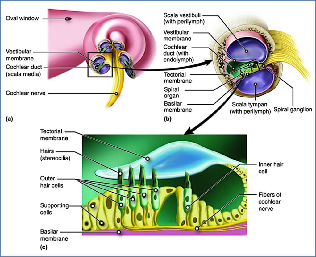
Inner Ear (Fluid-Filled Labyrinth)

- Cochlea (Hearing): Contains 3 ducts:
- Scala vestibuli (perilymph)
- Scala media (endolymph)
- Scala tympani (perilymph)

- Spiral Organ of Corti (on basilar membrane):
- Inner hair cells: Main sensory transducers
- Outer hair cells: Amplify sound signals
- Covered by tectorial membrane
Audiotransduction (How We Hear)
- Soundwaves → funnelled by auricle → external canal
- Vibrate tympanic membrane
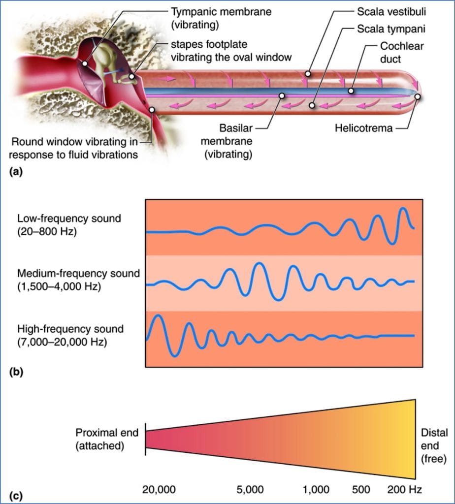
- Transferred via ossicles → oval window
- Oval window movement → perilymph wave in scala vestibuli
- Vibration passes through vestibular membrane → scala media
- High pitch = near oval window
- Low pitch = further along cochlea
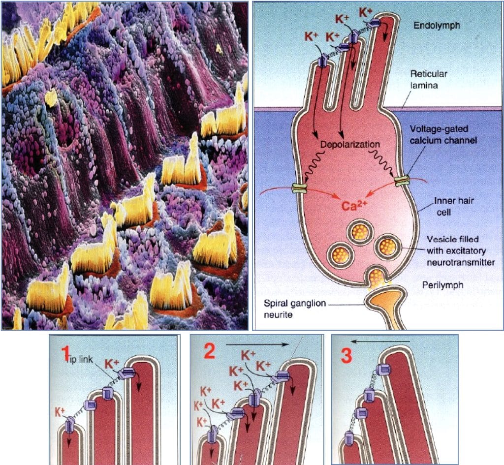
- Basilar membrane vibrates → hair cells bend into tectorial membrane
- Tip links stretch → K⁺ influx → depolarisation → Ca²⁺-mediated NT release
- Signal travels via cochlear nerve → brain
Hearing Pathway
Hair cells → Cochlear Nerve → Cochlear Nuclei (Medulla) →
→ Superior Olivary Nucleus → Lateral Lemniscus →
→ Inferior Colliculus → Medial Geniculate Body (Thalamus) →
→ Primary Auditory Cortex (Temporal Lobe)

Pitch & Volume
- Pitch: Coded by where along the basilar membrane the vibration occurs
- Volume: Coded by degree of hair cell displacement
Types of Deafness
- Conduction Deafness:
- Impaired transmission (e.g. wax, perforated tympanum, ossicle fusion)
- Sensorineural Deafness:
- Impaired transduction or nerve pathway (e.g. hair cell loss, CN VIII damage)
Equilibrium – Vestibular System
Vestibular Apparatus (Inner Ear)
- Vestibule:
- Utricle & Saccule → sense linear acceleration & gravity
- Contain Maculae (with otolithic membrane & CaCO₃ otoliths)

- Semicircular Canals:
- Detect rotational (angular) acceleration
- Each ends in ampulla containing Crista Ampullaris
- Hair cells project into cupula (gelatinous cap)
- Movement of endolymph bends cupula → signal generated
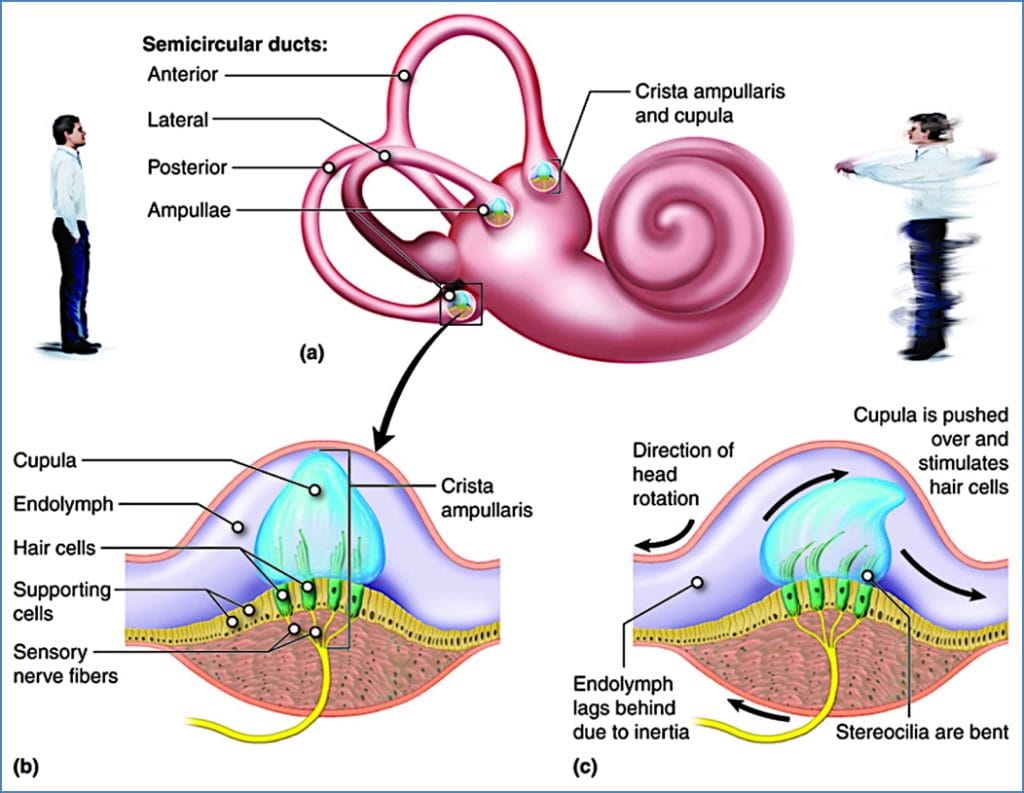
Planes of Detection:
- Anterior canal → ear-to-shoulder
- Posterior canal → nodding motion
- Lateral canal → shaking head ‘no’

Vestibular Pathway
- Hair cells → Vestibular Nerve → Vestibular Nuclei (Brainstem)
- Integrated with visual and somatic input
- Output to cerebellum, eye movement centres, and postural reflexes
Hair Cell Function
- Hair cells = mechanoreceptors
- Stereocilia movement affects tip link-gated K⁺ channels:
- Stretch = channel opens → depolarisation
- Compress = channel closes → hyperpolarisation
- NT release via Ca²⁺-mediated exocytosis
Summary – Hearing and Equilibrium
Hearing and balance are mediated by hair cells in the inner ear, which detect sound vibrations and head movements, respectively. The cochlea is responsible for hearing via the organ of Corti, while the vestibular apparatus regulates equilibrium through the maculae and semicircular canals. Understanding these pathways is crucial for diagnosing causes of deafness, vertigo, and gait disorders.
For a broader context, see our Nervous System Overview page.