Table of Contents
Overview – Brain Surface Anatomy
Surface anatomy of the brain refers to the visible landmarks observed on dorsal, ventral, medial, and coronal sections of the brain. Understanding these anatomical features is essential for localising functional regions, interpreting neuroimaging, and correlating clinical signs with neuroanatomical deficits. The brain’s surface is divided by prominent fissures and sulci into distinct lobes, each with specialised functions, while ventral and medial features reveal key structures in olfaction, vision, coordination, and homeostatic regulation.
Dorsal Landmarks
Major Fissures
- Longitudinal Fissure
- Separates left and right cerebral hemispheres
- Transverse Cerebral Fissure
- Separates occipital lobe from the cerebellum
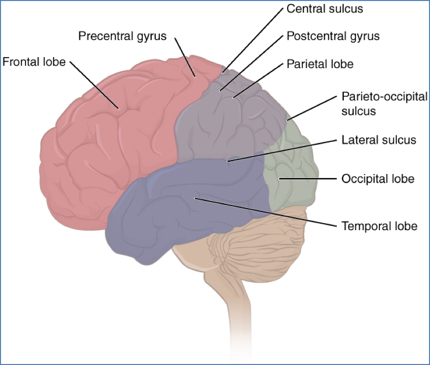
Prominent Sulci
- Central Sulcus
- Divides the frontal and parietal lobes
- Lateral Sulcus (Sylvian fissure)
- Separates the temporal lobe from the frontal and parietal lobes
- Parieto-Occipital Sulcus
- Divides the parietal lobe from the occipital lobe
Lobes of the Brain
- Frontal Lobe
- Anterior-most; involved in motor function, decision-making, personality
- Parietal Lobe
- Superior posterior; sensory integration
- Temporal Lobe
- Lateral location; auditory processing and memory
- Occipital Lobe
- Most caudal; primary visual processing centre
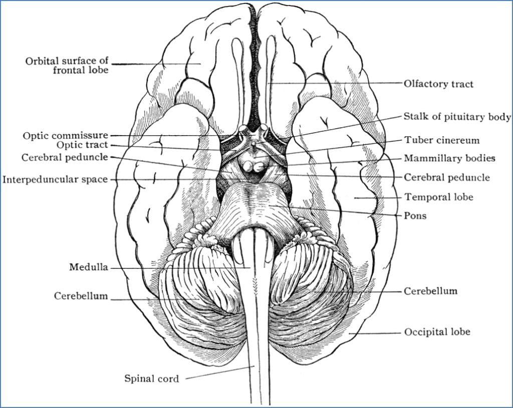
Ventral Landmarks
- Olfactory Bulbs
- Sense of smell; sit on the cribriform plate
- Optic Chiasm
- X-shaped crossover of optic nerves; important in visual field processing
- Infundibulum
- Connects the hypothalamus to the pituitary gland
- Hypothalamus
- Regulates autonomic functions: temperature, thirst, hunger
- Pituitary Gland
- Key endocrine gland for hormonal control
- Mammillary Bodies
- Limbic system component; involved in recollective memory
- Pyramids (Pyramidal Tracts)
- Located on medulla; carry corticospinal motor fibres
Medial Landmarks (Sagittal Section)
- Cingulate Gyrus
- Limbic structure above corpus callosum; regulates emotion and behaviour
- Corpus Callosum
- Large white matter tract connecting left and right hemispheres
- Lateral Ventricles
- Contain cerebrospinal fluid (CSF); lateral to midline
- Pineal Body (Gland)
- Regulates circadian rhythms (day/night cycle)
- Thalamus
- Relay station for sensory and motor signals; consciousness regulation
- Hypothalamus
- Integrative centre for homeostasis, endocrine control
- Pituitary Gland
- Controls metabolism, growth, sexual function, BP
- Colliculi (Tectum of Midbrain)
- Superior Colliculi: Visual reflexes, eye movement
- Inferior Colliculi: Auditory reflexes
- Cerebellum
- Coordinates voluntary movements, balance, and posture
- Pons
- Part of brainstem; regulates breathing and relays signals
- Medulla Oblongata
- Controls heart rate, respiratory rate, and reflex arcs
- Fourth Ventricle
- Contains CSF; lies between brainstem and cerebellum

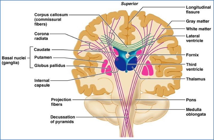
Coronal Section Landmarks
- Cortex (Grey Matter)
- Outer layer of brain; involved in conscious thought, memory, language, sensory and motor integration
- White Matter
- Myelinated axons that transmit signals to and from cortical areas
- Lateral Ventricles
- Bilateral CSF-filled cavities
- Caudate Nucleus
- Involved in motor planning, reward, memory, and emotion
- Corpus Striatum
- Part of basal ganglia; important in reinforcement learning and motor control
- Thalamus
- Sensory relay station for nearly all inputs to cortex
- Massa Intermedia
- Midline bridge connecting both thalami (in some individuals)
- Hippocampus
- Critical for memory consolidation and spatial navigation
Summary – Brain Surface Anatomy
Surface brain anatomy is defined by distinctive fissures, sulci, and lobes that demarcate functional regions. Dorsal, ventral, medial, and coronal views each reveal key structures responsible for motor, sensory, endocrine, emotional, and autonomic functions. This anatomical knowledge is fundamental to interpreting clinical signs and radiological imaging in neurology. For a broader context, see our Nervous System Overview page.