Table of Contents
Overview – Autonomic Nervous System Anatomy
Components of the autonomic nervous system anatomy (ANS) control involuntary visceral functions such as heart rate, digestion, and pupil size. The ANS consists of two main divisions—the sympathetic and parasympathetic nervous systems—which innervate the same organs but typically produce opposing effects. This dual innervation helps maintain physiological homeostasis during both rest and activity.
Divisions of the ANS
Sympathetic Nervous System
- “Fight or Flight”
- Mobilises body systems during activity
- Effects are widespread and longer-lasting
Parasympathetic Nervous System
- “Rest and Digest”
- Promotes maintenance and conserves energy
- Effects are localized and short-lived
Short duration is due to rapid breakdown of acetylcholine
ANS Control of Organs
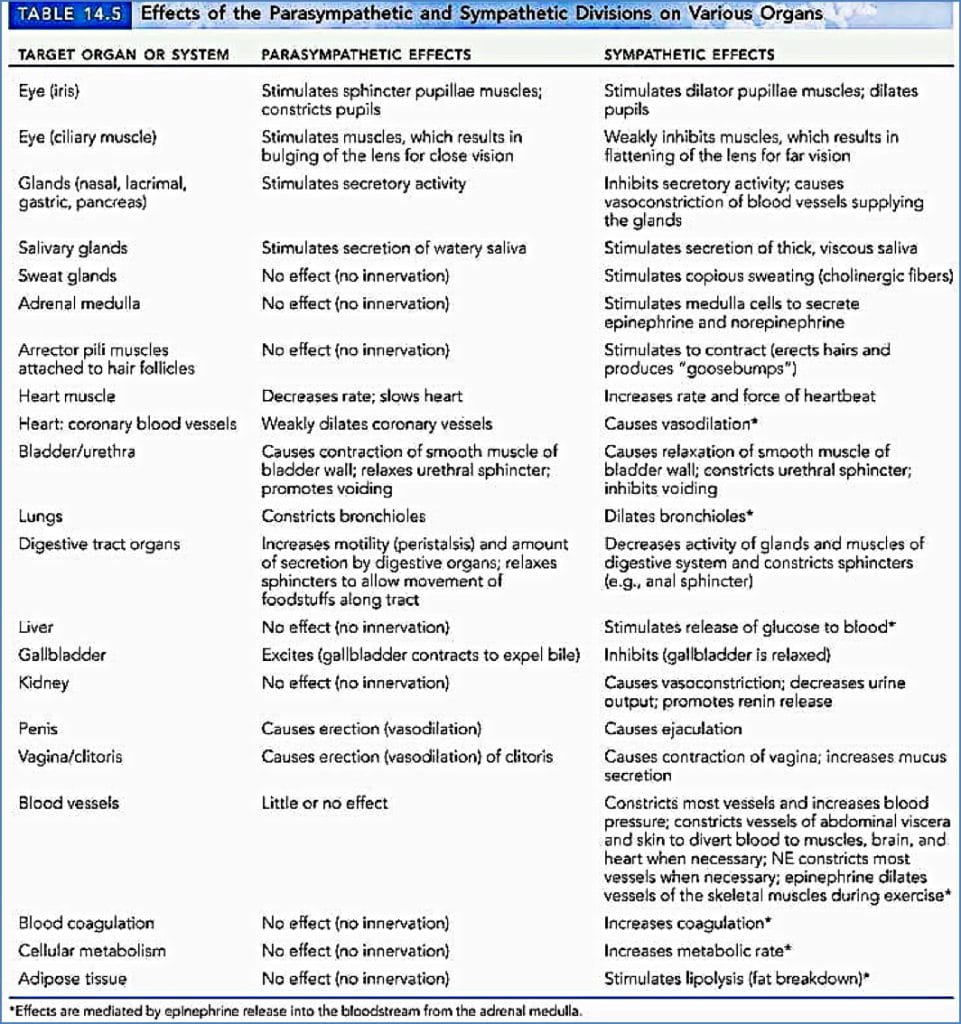
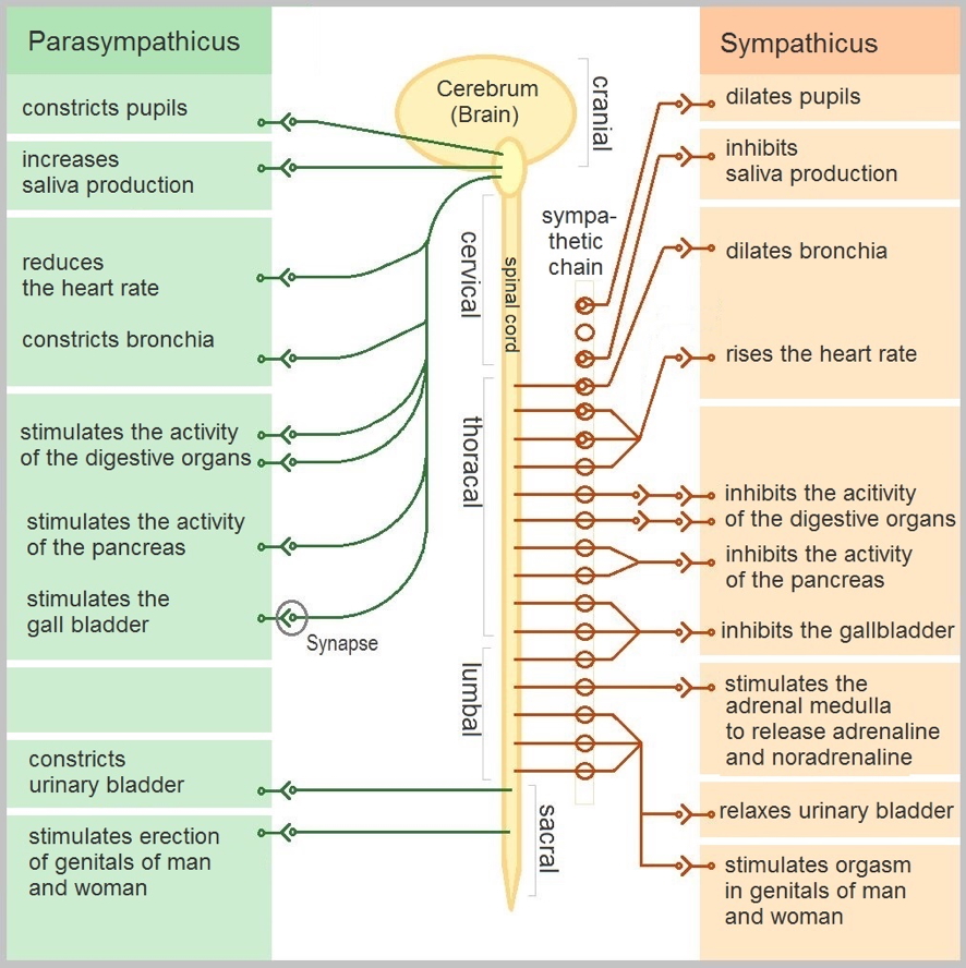
See the high-yield comparison table below for parasympathetic vs sympathetic effects across multiple organ systems (e.g. pupils, heart, lungs, digestive tract, etc.).
- Sympathetic effects are generally excitatory or mobilising
- Parasympathetic effects are inhibitory or restorative
Efferent Pathways & Ganglia
Unlike the somatic nervous system, which uses a single motor neuron, the ANS employs a two-neuron chain:
1. Preganglionic Neuron
- Cell body in the brainstem or spinal cord
- Axon synapses in an autonomic ganglion
2. Ganglionic (Postganglionic) Neuron
- Cell body in the autonomic ganglion
- Axon extends to the effector organ
Anatomical Differences Between Divisions
| Feature | Parasympathetic | Sympathetic |
|---|---|---|
| Origin | Craniosacral (Brainstem & S2–S4) | Thoracolumbar (T1–L2 spinal segments) |
| Fibre Lengths | Long preganglionic, short postganglionic | Short preganglionic, long postganglionic |
| Ganglia Location | Near/in effector organs (terminal ganglia) | In sympathetic trunks (near spinal cord) |
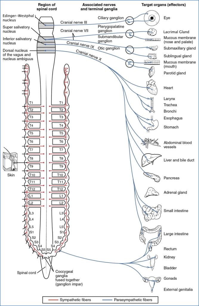
Parasympathetic Division (Craniosacral Outflow)
Origins
- Brainstem: CN III, VII, IX, X
- Sacral spinal cord: S2–S4
Cranial Outflow
- CN III (Oculomotor) → Pupil constriction, lens accommodation
- CN VII (Facial) → Stimulates lacrimal and salivary glands
- CN IX (Glossopharyngeal) → Stimulates parotid gland
- CN X (Vagus) → Thoracic & upper abdominal organs (heart, lungs, GI)
Sacral Outflow
- Originates from S2–S4
- Travels via pelvic splanchnic nerves
- Innervates distal large intestine, bladder, urethra, reproductive organs
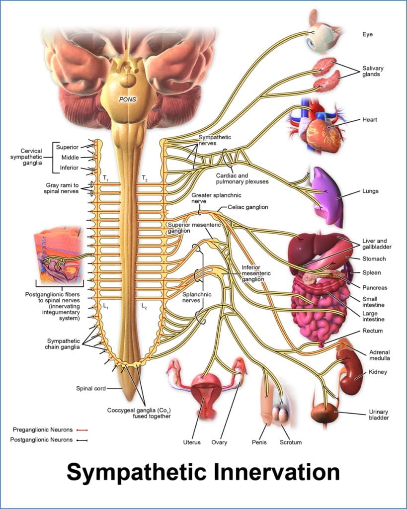
Sympathetic Division (Thoracolumbar Outflow)
Origins
- Preganglionic neurons in lateral horn of T1–L2 spinal cord segments
Pathway
- Preganglionic fibres exit via ventral root
- Enter white rami communicantes
- Synapse in sympathetic trunk ganglia
- May ascend, descend, or stay at same level
- Postganglionic fibres exit via gray rami communicantes
- Join spinal nerve → target organ
Rami colour indicates myelination:
- White rami: myelinated preganglionic fibres
- Gray rami: unmyelinated postganglionic fibres
Ganglia
- Located in paired sympathetic trunks flanking the vertebral column
Note: Although the sympathetic chain extends the spinal length, fibres only originate from T1–L2

Summary – Autonomic Nervous System Anatomy
The autonomic nervous system is divided into sympathetic (fight/flight) and parasympathetic (rest/digest) branches. Each uses a two-neuron chain and targets smooth muscle, cardiac muscle, and glands. Their opposing actions create balance and adaptability across physiological systems. For a broader context, see our Nervous System Overview page.