Table of Contents
Overview – Muscle Tissue
Muscle tissue is essential for producing movement, maintaining posture, stabilising joints, and generating heat. It exists in three primary forms—skeletal, cardiac, and smooth—each with specialised structure, function, and control mechanisms. Understanding muscle organisation, morphology, and contraction processes is foundational to mastering both physiology and clinical conditions affecting movement and coordination.
Types of Muscle Tissue
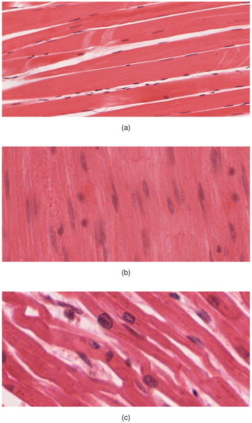
a) Skeletal Muscle
- Attached to bones for voluntary movement
- Long, cylindrical, multinucleated fibres
- Striated appearance due to sarcomeres
- Controlled by the somatic nervous system
b) Smooth Muscle
- Located in walls of visceral organs (e.g. gastrointestinal tract, urinary tract, uterus)
- Spindle-shaped, centrally located nuclei
- Lacks striations (no sarcomeres)
- Arranged in sheets, often in perpendicular layers
- Involuntary control via autonomic nervous system
c) Cardiac Muscle
- Found only in the heart
- Long, branched, cylindrical cells
- Single nucleus per cell
- Striated with sarcomeres
- Connected by intercalated discs:
- Mechanical coupling (desmosomes)
- Electrical coupling (gap junctions) → synchronised contraction
- Involuntary; controlled by autonomic nervous system
Functions of Muscle Tissue
- Produces skeletal movement
- Circulates blood via cardiac muscle
- Propels content through hollow organs (e.g. GI tract)
- Assists glandular secretion
- Maintains posture and structural support
Muscle Nomenclature
- Origin: Proximal attachment to immobile bone
- Insertion: Distal attachment to movable bone
- Prime Action: Movement of insertion while origin remains stationary (e.g. bicep curl)
- Reverse Action: Movement of origin while insertion is fixed (e.g. chin-up)

Muscle Morphology
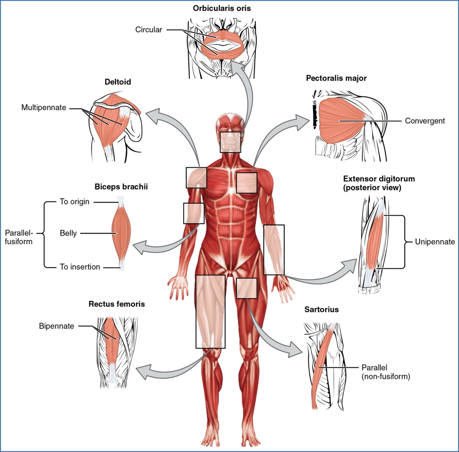
Fascicle Arrangements
- Parallel:
- Fascicles run parallel to long axis
- Large range of motion, less power
- Can be strap-like or fusiform
- Pennate:
- Fibres attach obliquely to a central tendon
- High power, less range
- Uni-, bi-, or multipennate
- Convergent:
- Broad origin, fascicles converge to single tendon
- Balanced power and range
- Circular:
- Fascicles arranged in concentric rings
- Found around external openings (e.g. mouth, eyes, anal sphincter)
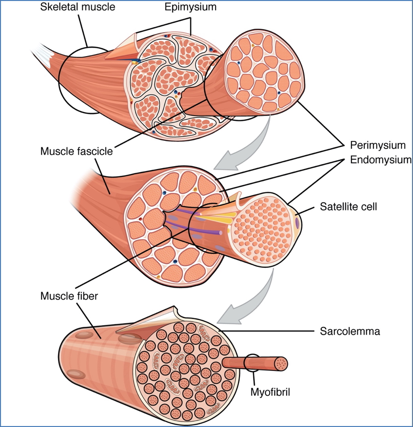
Organisation of Muscle Tissue
- Muscle Fibre: One contractile cell
- Contains multiple myofibrils, made of myofilaments (actin and myosin)
- Endomysium: Connective tissue around each fibre
- Fascicle: Bundle of muscle fibres
- Perimysium: Connective tissue around fascicles
- Whole Muscle: Encased by epimysium
- Tendon: Fusion of all layers; connects muscle to bone
Internal Machinery of Skeletal Muscle
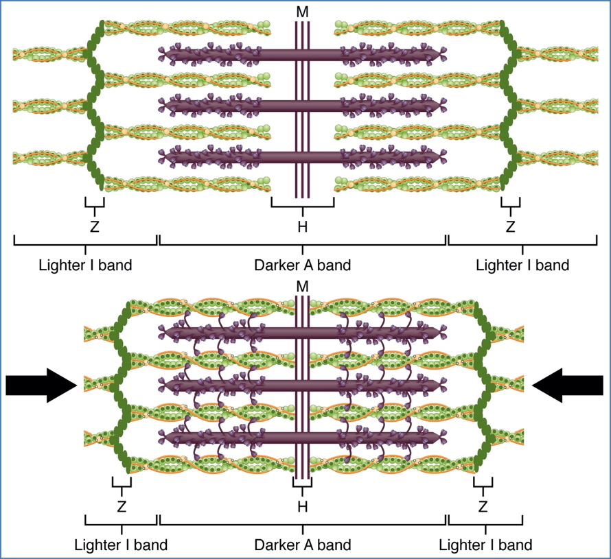
Sarcomere – Functional Unit
- Contains:
- Actin (thin filaments):
- Intertwined G-actin strands
- Tropomyosin blocks binding sites
- Troponin binds Ca²⁺ and controls tropomyosin
- Myosin (thick filaments):
- Long rodlike tails
- Heads form cross-bridges with actin during contraction
- ATPase activity for energy
- Actin (thin filaments):

Contraction Mechanism
- In relaxed state: minimal overlap of filaments
- Upon stimulation:
- Myosin heads bind actin → cross-bridge cycling
- Pulls actin toward centre of sarcomere
- Z-lines anchor sarcomeres, enabling coordinated contraction
Cellular Components of Muscle Fibres
- Sarcolemma: Plasma membrane
- Invaginates into T-tubules
- Conducts impulses deep into the fibre
- Sarcoplasm: Cytoplasm
- Rich in glycogen and myoglobin (O₂ storage)
- Sarcoplasmic Reticulum (SR):
- Surrounds each myofibril
- Stores and releases Ca²⁺
- Terminal cisternae flank each T-tubule
- Together form a triad at every I-band–A-band junction
- Mitochondria: Abundant to meet energy demands

Summary – Muscle Tissue
Muscle tissue is vital for voluntary and involuntary movements, structural support, and organ function. Its three types—skeletal, cardiac, and smooth—each possess unique structural and control features. Understanding fascicle arrangement, cellular architecture, and contraction mechanics is essential for diagnosing and managing neuromuscular conditions. For a broader context, see our Musculoskeletal Overview page.