Table of Contents
Overview – The Respiratory Examination
The respiratory examination is a fundamental clinical skill used to assess for pulmonary disease, respiratory failure, and systemic signs of underlying lung pathology. This exam integrates observation, palpation, percussion, and auscultation to evaluate respiratory mechanics and detect pathologies such as pneumonia, chronic obstructive pulmonary disease (COPD), asthma, pleural effusion, and lung cancer. Recognising signs like cyanosis, clubbing, tracheal deviation, and abnormal breath sounds is vital in diagnosing and managing respiratory conditions effectively.
Preparation
- Introduce yourself, explain the procedure, gain consent
- Wash hands and ensure privacy
- Position the patient: Sitting upright, fully exposed from waist up
General Inspection
- Alertness and Orientation
- Signs of Respiratory Distress:
- Tripoding
- Pursed-lip breathing (COPD)
- Intercostal recession/tracheal tug (restrictive disease)
- Audible wheeze, cough, or stridor
- Body Habitus:
- Obesity (blue bloaters – chronic bronchitis phenotype of COPD)
- Cachexia (pink puffers – emphysema or malignancy)
- Chest wall deformities: pectus excavatum, carinatum, barrel chest
- Colour:
- Cyanosis
- Plethora (polycythaemia)
- Pallor (anaemia)

Vital Signs
- Pulse: Tachycardia → anaemia, infection, hypoxia
- Blood Pressure:
- Normal or postural variation (severe anaemia)
- Pulsus paradoxus (COPD, asthma, tamponade, pneumothorax)
- Respiratory Rate: Usually elevated in lung disease
- SpO₂: May be preserved or reduced; note if on supplemental oxygen
- Temperature: Fever suggests infection
Hands
- Capillary refill time (CRT)
- Clubbing: Chronic hypoxia, bronchiectasis, lung cancer
- Pale nails and koilonychia: Iron-deficiency anaemia
- Palmar crease pallor
- Tar staining: Long-term smoking
- Muscle wasting/weakness: Pancoast tumour (intrinsic hand muscles)
- Asterixis: CO₂ retention
Arms
- Hypertrophic pulmonary osteoarthropathy (HPOA): Lung cancer
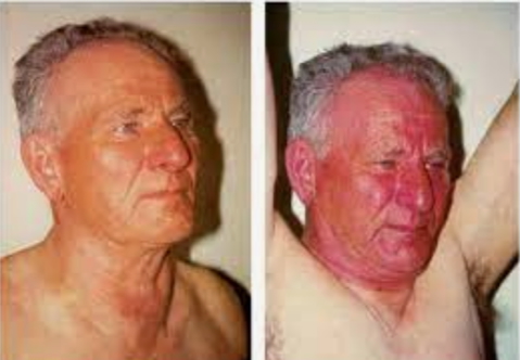
- Pemberton’s sign: SVC obstruction → facial plethora with raised arms
Face
- Plethora: Polycythaemia
- Skin lesions: Basal or squamous cell carcinoma
- Conjunctival pallor, subconjunctival haemorrhages (coughing)
- Glossitis/Stomatitis: Anaemia
- Cyanosis: Central or peripheral
- Dental signs: Tar-stained teeth (smoking)
- Leukoplakia/Erythroplakia: Premalignant lesions from smoking
- Horner’s Syndrome: Ptosis, miosis, anhydrosis → Pancoast tumour
Neck
- Lymphadenopathy: Infection, malignancy
- Virchow’s node: Supraclavicular node suggestive of intrathoracic malignancy
- Raised JVP: Pulmonary hypertension, SVC obstruction
- Abdominojugular reflux: Negative in SVC obstruction
- Tracheal deviation: Tension pneumothorax, atelectasis
- Tracheal tug: Restrictive lung disease
Chest
Inspection (from front and behind)
- Scars, deformities
- Symmetrical chest movement
- Use of accessory muscles
Palpation
- Chest expansion: Reduced asymmetrically in collapse or effusion
- Tactile fremitus: ↑ in consolidation
- Hoover’s sign: Inward movement of lower ribs (severe COPD)
- Chest wall tenderness: Fracture, tumour
Percussion
- Dullness: Consolidation, collapse
- Stony dullness: Pleural effusion
- Hyper-resonance: Pneumothorax, asthma, emphysema
- Lower border: 6th rib anteriorly = normal
Auscultation
- Breath Sounds:
- Vesicular = normal
- Bronchial = consolidation
- Diminished = effusion, pneumothorax
- Crackles = pulmonary oedema, pneumonia
- Wheeze = asthma, COPD
- Pleural rub = mesothelioma, pleuritis
- Vocal Resonance: Increased in consolidation
Always offer a cardiovascular exam to complete the assessment.
Abdomen
- Liver percussion: Displacement (ptosis) from lung hyperinflation
- Hepatomegaly: Right heart failure
Legs
- Oedema: Cor pulmonale, right heart failure
- Calf tenderness: Deep vein thrombosis → risk of PE
- Signs of venous stasis: Ulcers, shiny skin, hair loss
Feet
- Capillary refill & perfusion
- Clubbing, koilonychia, pale nails (as above)
Summary – The Respiratory Examination
The respiratory examination systematically assesses for signs of respiratory distress, obstructive and restrictive lung diseases, and systemic manifestations of malignancy or infection. Recognising patterns like tracheal deviation, tactile fremitus changes, and abnormal breath sounds can significantly narrow differential diagnoses. For a broader context, see our Clinical Skills Overview page.