Table of Contents
Overview – The Haematological Examination
The haematological examination is a comprehensive clinical skill that helps detect signs of anaemia, blood cancers, clotting disorders, and systemic haematological conditions. It involves inspection for pallor, lymphadenopathy, hepatosplenomegaly, bleeding tendencies, and systemic manifestations of blood dyscrasias. The haematological examination is essential in identifying red flag signs of malignancy, iron deficiency, haemolysis, and bone marrow suppression.
General Inspection
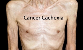
- Wasting/Cachexia → Malignancy
- Ethnic Features:
- Mediterranean background with frontal bossing → Thalassaemia
- African descent → Sickle cell anaemia
- Skin and Colour:
- Gross pallor → Anaemia, haematological malignancies
- Jaundice with scratch marks → Haemolytic anaemia
- Cyanosis
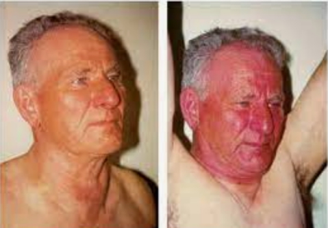
- Facial plethora → Polycythaemia, superior vena cava (SVC) obstruction (e.g. from lymphoma)
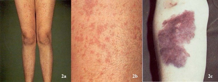
- Bruising, petechiae, purpura → Thrombocytopenia, leukaemia, haemophilia
- Lymphadenopathy or Neck Masses → Lymphoma or other haematological malignancies
- Abdominal Distension → Splenomegaly/hepatomegaly in haematological disease
Vital Signs
- Pulse:
- Tachycardia → Anaemia, infection
- Blood Pressure:
- Postural hypotension → Anaemia
- Respiratory Rate:
- Tachypnoea → Anaemia
- Temperature:
- Fever → Infection or haematological malignancy
Hands
- Perfusion:
- Warm, well perfused → Normal
- Cool peripheries, prolonged capillary refill time → Shock or hypovolaemia
- Nails:
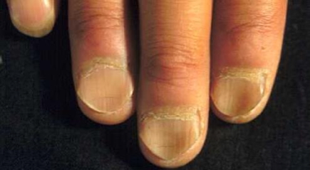
- Koilonychia (spoon nails) and pale nails → Iron deficiency anaemia
- Blue lunulae → Wilson’s disease

- Palms:
- Pallor → Anaemia
- Erythema → Polycythaemia, chronic leukaemia
- Other Signs:
- Peripheral cyanosis
- Digital infarcts or Raynaud’s phenomenon → Multiple myeloma, autoimmune diseases
- Arthropathy → Haemophilia, connective tissue disease
- Gouty tophi → Increased cell turnover (e.g. myeloproliferative disease)
- Dupuytren’s contracture → Megaloblastic anaemia, alcoholism
Arms
- Bruising, Petechiae, Purpura → Platelet or coagulation disorders
- Scratch marks → Haemolytic jaundice
- Lymph Nodes:
- Epitrochlear, axillary, infraclavicular, supraclavicular, subpectoral, lateral, central
- Examine for size, consistency, tenderness, mobility → Lymphoma vs infection
Face
- Eyes:
- Conjunctival pallor → Anaemia
- Scleral icterus → Haemolysis
- Oral Cavity:
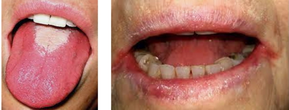
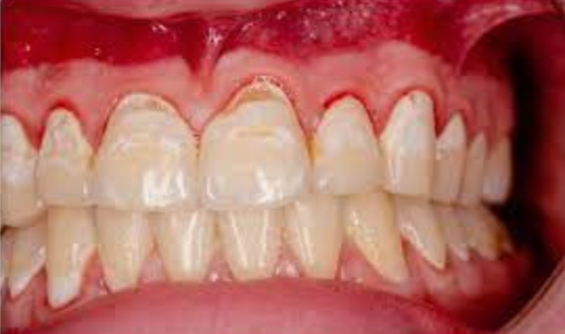
- Angular stomatitis, glossitis → Anaemia (especially B12/folate)
- Buccal petechiae → Thrombocytopenia or leukaemia
- Candidiasis → Immunosuppression
- Gum hypertrophy → Acute myeloid leukaemia (AML) or methotrexate treatment
- Tonsillar enlargement → MALT lymphoma
- Facial Plethora → Polycythaemia
- Pemberton’s sign → SVC obstruction from mediastinal mass
Neck
- Lymph Nodes:
- Submental, submandibular, pre/post-auricular, jugular chain, occipital, posterior triangle
- Characteristics:
- Rubbery, firm, non-tender → Lymphoma
- Hard and immobile → Carcinoma
- Tender, mobile → Infection
- Other Findings:
- Neck swellings
- Check for dysphagia or odynophagia → Plummer-Vinson syndrome (oesophageal web in iron deficiency)
- Thyroid enlargement → Hypothyroidism leading to menorrhagia and secondary anaemia
- Radiotherapy tattoos
- Basal cell carcinoma or squamous cell carcinoma → From immunosuppression
Chest
- Skin:
- Bruising, petechiae, purpura → Coagulopathies
- Infections → Common due to immunosuppression
- Heart Sounds:
- Systolic flow murmur → Severe anaemia
- Bony Tenderness:
- Ribs, clavicles, spine, hips → Haematological malignancies (e.g. myeloma, leukaemia)
Abdomen
- Skin:
- Bruising, petechiae, purpura
- Palpation:
- Hepatomegaly and splenomegaly → Common in leukaemia, lymphoma
- Para-aortic lymphadenopathy
- Ascites
- Kidney enlargement → Anaemia of chronic kidney disease
- Rectal:
- Blood per rectum → Leukaemia or other causes
Inguinal
- Lymph Nodes → Haematological malignancies
- Testicular Masses in Children → Acute lymphoblastic leukaemia (ALL)
- Digital Rectal Examination → Assess for rectal lymphoma
Legs
- Bruising, Petechiae, Purpura
- Scratch Marks → Haemolytic jaundice
Feet
- Peripheral Perfusion & CRT
- Koilonychia, Pale Nails → Iron deficiency anaemia
- Sole Erythema → Polycythaemia
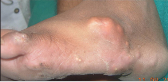
- Gouty Tophi → Myeloproliferative disorders
Summary – The Haematological Examination
The haematological examination identifies signs of anaemia, clotting disorders, leukaemia, lymphoma, and other haematological malignancies. From koilonychia and lymphadenopathy to hepatosplenomegaly and facial plethora, it provides crucial insight into underlying systemic disease. For a broader context, see our Clinical Skills Overview page.